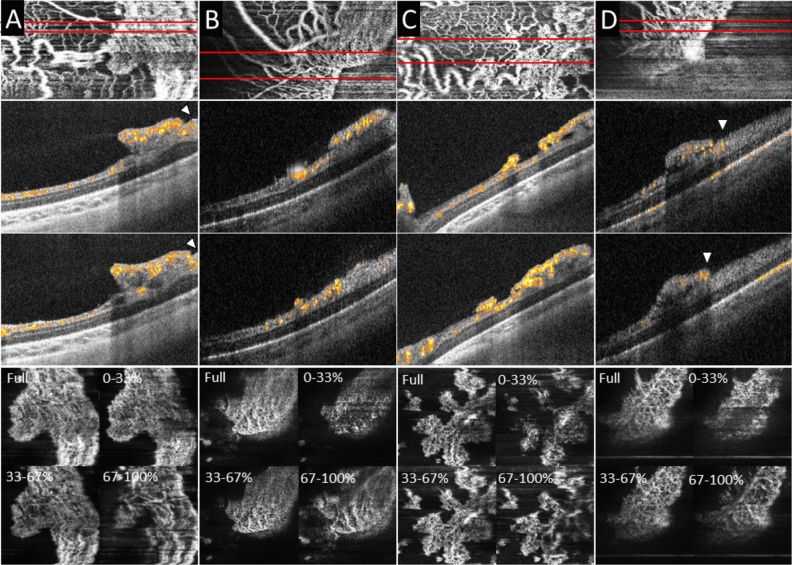
Figure 3.
Extraretinal neovascular plaques and distribution of vascular flow by axial location within the neovascular plaques on handheld optical coherence tomography angiography (OCTA). Top row: en face OCTA flow; Second and third rows: B-scans with flow overlay; Bottom row: en face sub-sectional flow (generated by the percentage of distance from the top to bottom of the neovascular plaque). In three eyes with treatment requiring-ROP, extraretinal neovascular plaques were found elevated from the retinal surface along the vascular-avascular junction. On OCT B-scans with flow overlay, the retinal vascular flow signal extends to the edge or beyond the edge of the neovascular plaque (arrowheads). The vascular flow signal on cross-section on B-scans with flow overlay and the sub-sectional flow analysis (Full, 0–33%, 33–67%, and 67–100%) showed finer capillaries at the surface of the plaque (vitreous side) and larger, more dilated capillaries at the bottom of the plaque (retina side).

