Fig. 2.
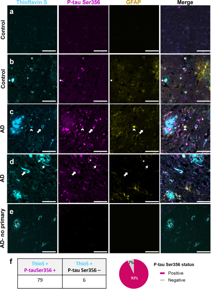
p-tau Ser356 is observed around plaques, in dystrophic neurites, neuropil threads and tangles in human postmortem brain. Paraffin sections of control (a, b) or AD (c–e) post-mortem brain stained for plaques/tangles (Thioflavin S—cyan), p-tau Ser356 (magenta) and reactive astrocytes (GFAP—yellow). In human brain sections, p-tau Ser356 (magenta) is found in neurofibrillary tangles (notched arrows), neuropil threads (thin arrows) and dystrophic neurites around plaques (asterisks). p-tau Ser356 is also observed in some GFAP (yellow) positive astrocytes (chevrons). Control brain with little/no Thioflavin S or p-tau Ser356 staining (a). Control brain with evidence of dystrophic neurites (b), AD brains with evidence of plaques (c, d), dystrophic neurites (c, d), neuropil threads (d) and tangles (c, d). No primary antibody control (e). Scale bar 50 μm. f Quantification of Thioflavin S, p-tau Ser356 double positive tangles, compared to Thioflavin S only tangles shows the vast majority of tangles counted in this study (93%) contain p-tau Ser356

