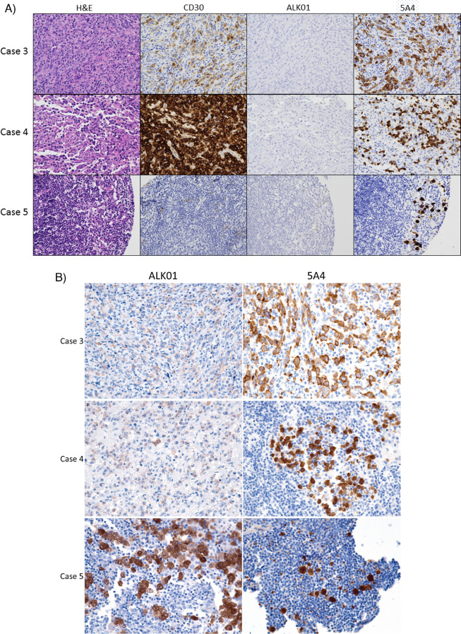
Fig. 1.
Cases that were classified as ALK-negative ALCL originally. A The images show tissue from the tissue microarrays of three cases that were initially classified as ALK-negative ALCL on the basis of a negative ALK01 stain but showed strong positivity with the 5A4 antibody. Rare dimly positive cells that could be misinterpreted as nonspecific staining were seen in the ALK01-stained samples. Case 5 showed rare dim CD30-positive cells and very rare dimly ALK01-positive cells. 5A4 brightly highlights rare cells. Images are × 400 magnification. B Whole tissue sections of cases 3, 4, and 5 were stained with the ALK01 and 5A4 protocols. In cases 3 and 4, the ALK01 protocol revealed ALK expression in the lymphoma cells, but the signal was notably dimmer than with the 5A4 protocol

