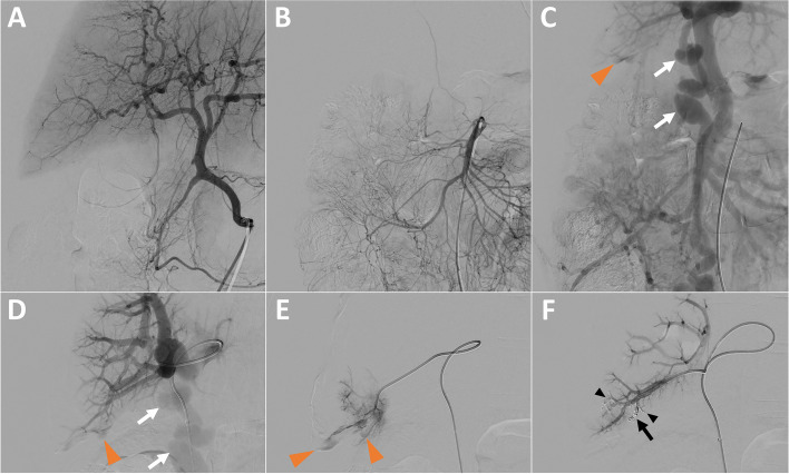
Fig. 2.
Portal Vein Embolization (PVE) after hepatic arteriography, guided by arterial portography. A Hepatic arteriography showing no arterial active bleeding. B & C Superior mesenteric arteriography (B) with subsequent arterial portography (C) showing active intraperitoneal bleeding of portal origin in segment VI (orange arrowhead) and a voluminous paraumbilical vein (white arrows). D Direct portography after ultrasound-guided puncture of the paraumbilical vein (white arrows) showing active portal bleeding in segment VI (orange arrowhead). E Catheterization of a distal portal branch in segment VI and injection of contrast agent to confirm the location of the bleeding (orange arrowheads). F Control portography, after embolization with 2 detachable microcoils (black arrow) and a glue/lipidol mixture (black arrowheads), confirming complete cessation of bleeding

