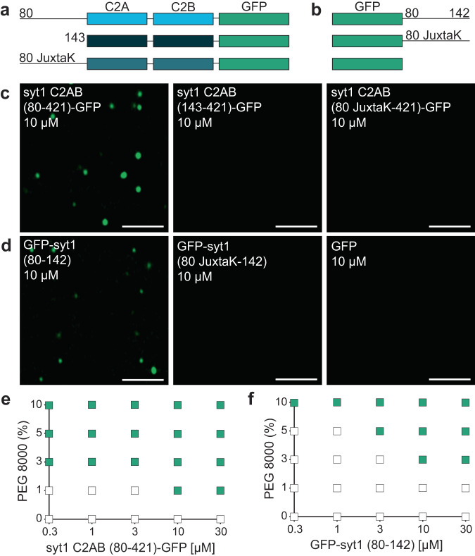
Fig. 3. The IDR region of syt1 is necessary and sufficient for LLPS.
Schematics of the syt1 C2AB-GFP a and isolated GFP-tagged juxtamembrane linker b fusion proteins used to study LLPS. c syt1 C2AB (80–421)-GFP and d GFP-syt1 (80–142) form droplets in a buffer of physiological salt and 3% PEG 8000, whereas syt1 C2AB lacking this linker, or in which the linker has been mutated (JuxtaK), fails to form droplets; n = 3. e, f Phase diagram of syt1 C2AB (80–421)-GFP and GFP-syt1 (80–142) with varying protein and PEG 8000 concentrations. The buffer condition was 25 mM Tris-HCl (pH 7.4), 100 mM NaCl. Green squares indicate the appearance of droplets, whereas white squares indicate no droplet formation; n = 3. (Scale bars, 3 µm).

