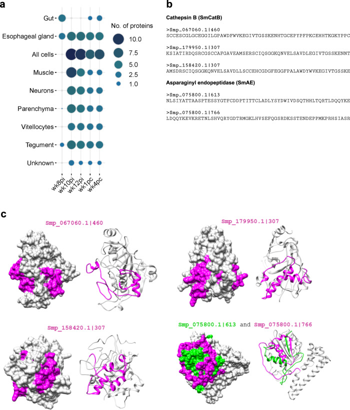
Fig. 4. Expression localization of S. mansoni extracellular proteins containing the peptides captured by antibodies from self-cured and challenge-resistant rhesus macaques.
a Bubble plot depicting the number of extracellular proteins comprising enriched peptides at predicted S. mansoni organ expression locations using publicly available single-cell data. Circle size and color range (legend on the right) represent the number of distinct proteins enriched in samples incubated with rhesus macaques’ plasma collected at each indicated week post-infection (wk8pi, wk10pi, wk12pi) or week post-challenge (wk1pc, wk4pc), or in samples incubated with hamsters’ sera collected at wk12pi or wk22pi, which are analyzed together here under the label “Hamster”. b The 58-mer enriched peptide sequences from three cathepsin B paralogs (Smp_067060.1|460, Smp_179950.1|3-7, Smp_158420.1|307) and two stretches from the asparaginyl endopeptidase (Smp_075800.1|613 and Smp_075800.1|766). These proteins are predicted as secreted in the parasite gut and begin to be recognized by the rhesus macaques’ plasma at wk8pi. c Homology models from predicted extracellular proteins expressed in the parasite gut highlighting enriched peptides on the specific protein surfaces. Protein structures are depicted in grey, while enriched peptides are color-coded in pink (representing most peptides) or green (specifically Smp_075800.1|613).

