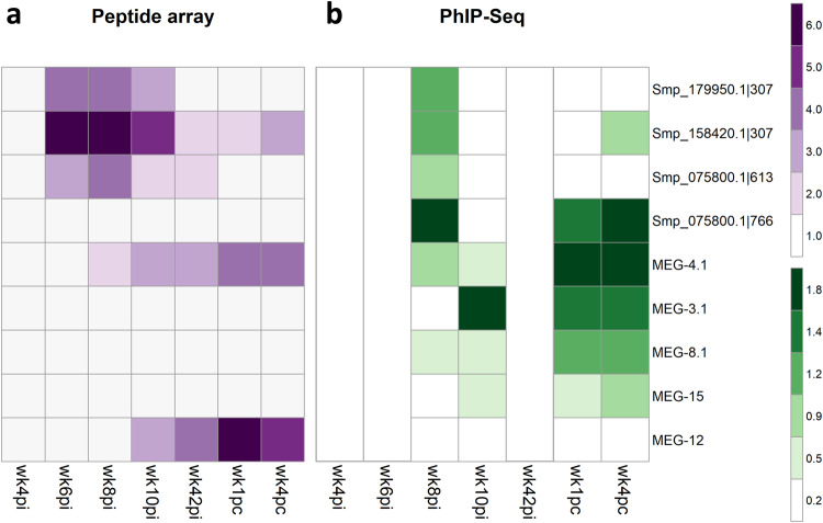
Fig. 6. Heatmap of significantly reactive 15-mer peptides recognized by antibodies in the plasma from rhesus macaques self-cured and challenge-resistant to S. mansoni.
Each line represents one peptide from a different protein or multiple peptides from the indicated MEG family member’s alternatively spliced MEG protein variants. These were selected for the peptide microarray based on their enriched signal in the PhIP-Seq analysis. a Median intensity signal in the peptide microarray among the different rhesus macaques and across the entire set of 15-mer peptides is shown in purple (see color scale) for those entries with significantly reactive peptide motifs, as determined with the pepStat tool. Columns represent the four time points at weeks post-infection (wk4pi, wk6pi, wk8pi, wk10pi), the time point at challenge (wk42pi), and the two time points at weeks post-challenge (wk1pc, and wk4pc). In addition, MEG-12, not enriched in the PhIP-Seq analysis, was included in the peptide microarray. b For comparison, the median enrichment values among the different rhesus macaques of significantly enriched peptides in the PhIP-Seq assay are shown in green (see color scale). Empty columns represent time points not assayed by PhIP-Seq.

