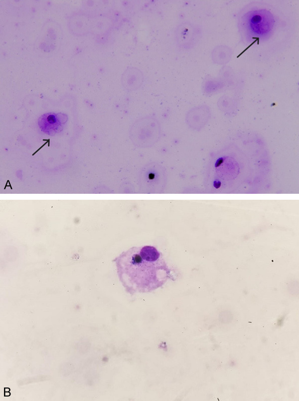
Figure 1.

A. Histiocytes that have phagocytosed erythrocytes in the CSF smear prepared by the cytocentrifuge method (arrows). B. In the CSF smear prepared by the cytocentrifuge method, erythrocytes and lymphocyte-phagocytosed histiocytes are observed (×1000 May Grünwald Giemsa staining).
