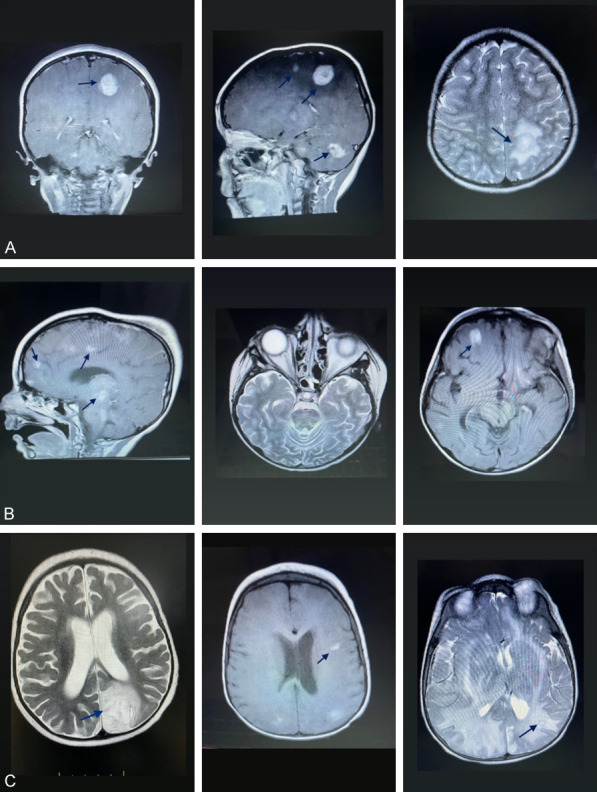
Figure 3.

A. MRI of patient 10 at the time of admission (MRI showing nodular enhancing lesions in the contrast-enhanced weighted-T1 sequence, and a peripherally hypointense and centrally hyperintense lesion in T2A). B. Control MRI of patient 10 (T1A showing contrast-enhancing lesions in the frontal lobe and brainstem). C. MRI of patient 11 at admission (T2A showing a hyperintense lesion located in the parieto-occipital cortical and subcortical areas. Fluid-attenuated inversion recovery (FLAIR) showing increased signal in bilateral parieto-occipital white matter and left parietal subcortical lesion) (arrows).
