
Figure 4.
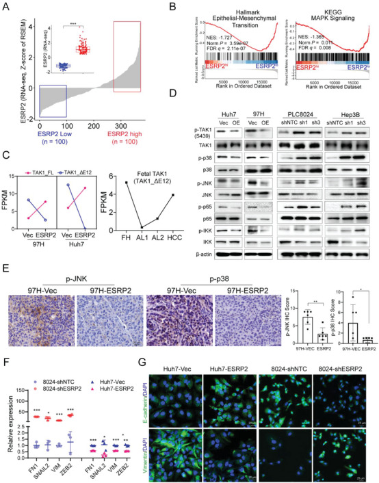
ESRP2 mediates inclusion of exon 12 in TAK1 pre‐mRNA and inactivates p38MAPK signaling. A) ESRP2 expression in HCC specimens (n = 371) based on TCGA datasets. Inset shows comparison between low (blue box, n = 100) and high (red box, n = 100) ESRP2 expression cohorts. RSEM, RNA‐Seq by Expectation Maximization. B) Representative gene sets enriched by GSEA analysis of DEGs from HCC specimens with low or high ESRP2 expression in the TCGA cohort. C) Left: FPKM expression values of TAK1 variants based on RNA‐Seq data from Vec‐ or ESRP2‐ transfected HCC cells; Right: FPKM expression values of the fetal TAK1 variant analyzed using transcriptomic data from fetal hepatocytes (FH), adult liver (AL) and HCC samples. D) Western blot analysis of phosphorylation of TAK1, p38, JNK, p65, IKK in ESRP2‐overexpressed or silenced HCC cells. E) Representative images of IHC staining for p‐JNK and p‐p38 in xenograft tumors induced by Vec‐ or ESRP2‐ transfected HCC cells. IHC scores were calculated and shown in the bar chart, with each dot representing an individual xenograft tumor. F) qRT‐PCR was used to assess the relative expression of EMT markers in ESRP2‐overexpressed or silenced HCC cells. G) Immunofluorescence staining of E‐cadherin and Vimentin in ESRP2‐overexpressed or silenced HCC cells. Cell nuclei were counterstained with DAPI (blue). Scale bars: 20 µm.