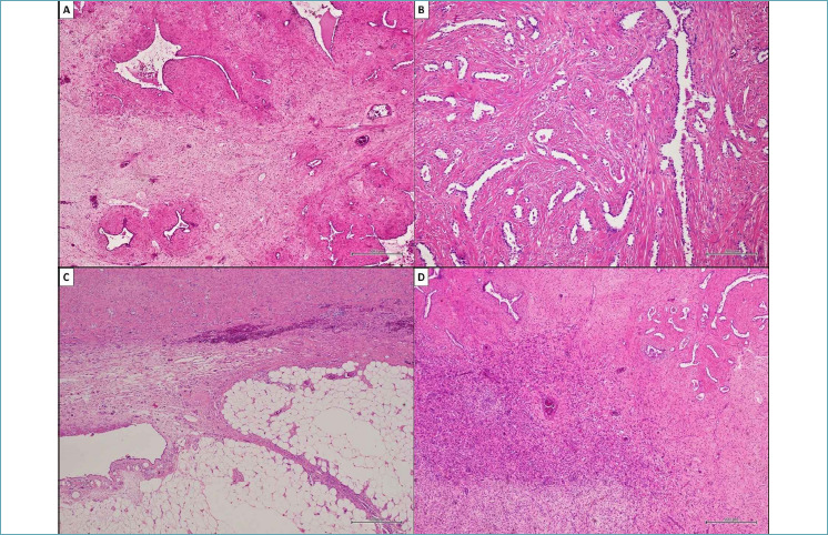
Figure 3.

Histological findings observed in the surgical specimen. (A) The tumor consisting of bland and monomorphic spindle cells. Neoplastic population showed a predominant fascicular organization and a typical zonal pattern, with hypercellular and hypocellular areas in which many tubular and cleft-like spaces of entrapped normal respiratory epithelium were involved. (B) Detail of tubular and cleft-like spaces of entrapped respiratory epithelium. (C) Scattered foci of adipose tissue were also observed between neoplastic cells. (D) Detail of hypercellular area composed of relatively bland and monomorphic spindle cells. (A, C, D) Hematoxylin and eosin, original magnifications x500. (B) Hematoxylin and eosin, original magnifications x200.
