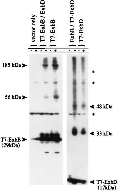
FIG. 3.
In vivo cross-linking of cells expressing T7-ExbB or T7-ExbD. ΔexbBD cells (KP1269) expressing plasmids pACYC184 and pET-24(a)+ (vector controls), pKP360 (ExbD), pKP339 (T7-ExbB), pKP361 (ExbB), or pKP323 (T7-ExbD) were untreated (−) or cross-linked in vivo with monomeric formaldehyde (+) as described in Materials and Methods. Samples representing equal cell numbers were electrophoresed on SDS–11% polyacrylamide gels, and T7 epitope-tagged proteins were detected by immunoblot analysis with an anti-T7 epitope tag monoclonal antibody. The small amount of T7-ExbB monomer signal detected in vector-only controls is due to bleed-through from the adjacent lane and is visible only after extended exposures. The bands denoted by an asterisk at 42, 85, and 120 kDa appear to arise from unidentified cross-reactive proteins since they appear in lanes carrying vector controls.

