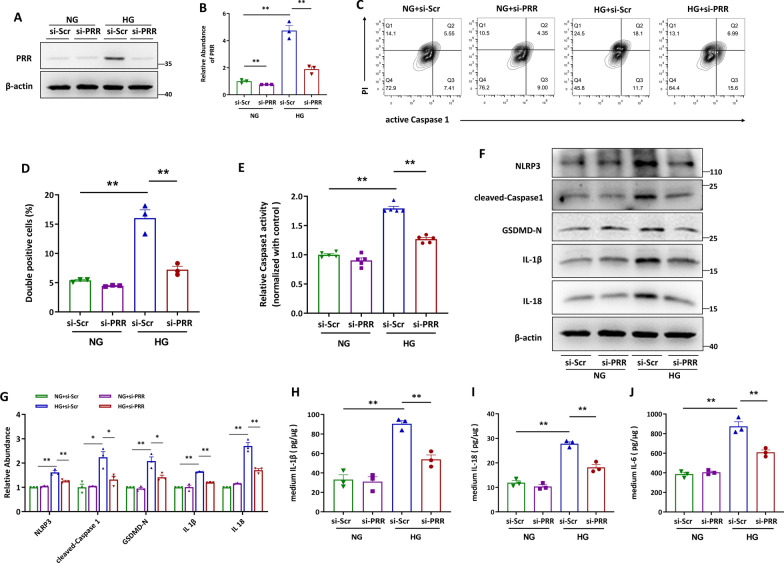
Fig. 2.
Knockdown of PRR effectively blocked high glucose (HG)-augmented HK-2 cell pyroptosis. A and B Western blot analyses (A) and quantitative data (B) showed that knocking down by siRNA diminished HG-induced PRR expression. (n = 3). C and D Flow cytometry analysis showed that HG induced active Caspase1+PI+ HK-2 cells were decreased by PRR siRNA (C) and quantitative data (D). (n = 3). E PRR was silenced in HG stimulated HK-2 cells, and the Caspase1 activity in cell lysis was determined by kits (n = 5). F and G Western blot analyses (F) and quantitative data (G) showed that PRR siRNA blocked HG-induced NLRP3, cleaved-Caspase1, GSDMD-N, IL-1β and IL-18 expression in HK-2 cells (n = 3). H and I PRR was ablated in HG treated HK-2 cells, and the IL-1β (H), IL-18 (I) or IL-6 (J) concentration in the culture medium was determined by ELISA, and then normalized by protein concentration in cell lysates (n = 3). Data are presented as mean ± SEM of biologically independent samples. ∗ P < 0.05, ∗∗ P < 0.01. One-way ANOVA was used to analyze the data among multiple groups, followed by Tukey’s post hoc test

