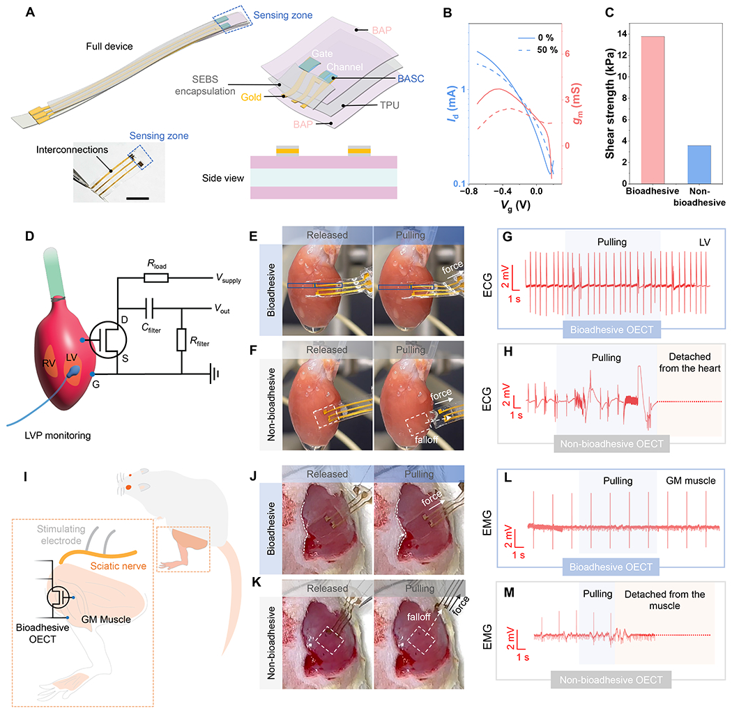
Fig. 5. Fully-bioadhesive OECT sensor and the use for ex vivo and in vivo electrophysiological recording.

(A) Device structure and picture. Scale bar: 5 mm. (B) Transfer curves for a fully-bioadhesive OECT under 0 % and 50 % strains. (C) Shear strength of the adhesion between a fully-bioadhesive OECT on the porcine muscle tissue, in comparison to an OECT with non-bioadhesive surface. (D) Schematic showing the use of the OECT sensor for ECG recording on a heart surface, and the circuit diagram. (E) Photographs showing a fully-bioadhesive OECT attached to an isolated rat heart surface maintaining stable contact during mechanical agitation. (F) Comparison of a non-bioadhesive OECT, for which capillary-based attachment cannot maintain conformable and stable contact. (G) ECG signals recorded by the fully-bioadhesive OECT on the LV. (H) ECG recording by the non-bioadhesive OECT, which ceased after the device detachment from the heart. (I) Schematic showing the use of the OECT sensor for EMG recording on the GM muscles upon stimulation of the sciatic nerve. (J) Photographs showing a fully-bioadhesive OECT attached to the GM muscle maintaining stable contact during mechanical agitation. (K) Comparison of a non-bioadhesive OECT, for which capillary-based attachment cannot maintain conformable and stable contact. (L) EMG signals recorded by the fully-bioadhesive OECT on the GM muscle. (M) EMG signals recorded by the non-bioadhesive OECT on the GM muscle, which ceased after the device detached from the muscles.
