Figure 1:
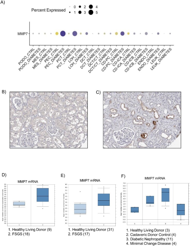
MMP-7 expression in human kidney diseases. (A) Human kidney single cell transcriptomics in control and diabetic kidney samples. Multiple cell types appear to express higher levels of MMP-7 mRNA in diabetic kidneys: PEC (parietal epithelial cells), PCT (proximal convoluted tubule), CD-PC (collecting duct principal cell), CD-ICB (collecting duct-intercalated cell type B) and endo (endothelium) [15]. https://humphreyslab.com/SingleCell/. Reproduced with permission from Prof. Ben Humphreys. (B, C) Human kidney immunohistochemistry in ProteinAtlas. Although both kidneys were classified as normal, panel A displayed back-to-back tubules, characteristic of healthy kidneys (male, age 16), while panel B shown widened interstitial spaces (female, age 59), characteristic of chronic kidney disease. Intense cytoplasmic staining is observed in distal tubules in panel B. https://www.proteinatlas.org/ENSG00000137673-MMP7/tissue/kidney#img. Image credit: Human Protein Atlas [16]. (D–F) MMP7 gene expression of micro-dissected tubulointerstitial samples from: (D) 18 focal segmental glomerulosclerosis (FSGS) patients and nine healthy living donors. Fold Change: 5.274; P‑value: 7.91E-5. ERCB Nephrotic Syndrome TubInt Dataset. http://v5.nephroseq.org/; (E) 17 FSGS patients and 31 healthy living donors. Fold change: 1.93; P‑value: 0.006. Ju CKD Tubint dataset [41]. http://v5.nephroseq.org/; (F) 3 living donors (controls), 4 minimal change disease (MCD), 4 cadaveric donors, and 11 diabetic nephropathies. Fold change Controls vs MCD: 1.09. Schmid Diabetes TubInt Dataset [42].

