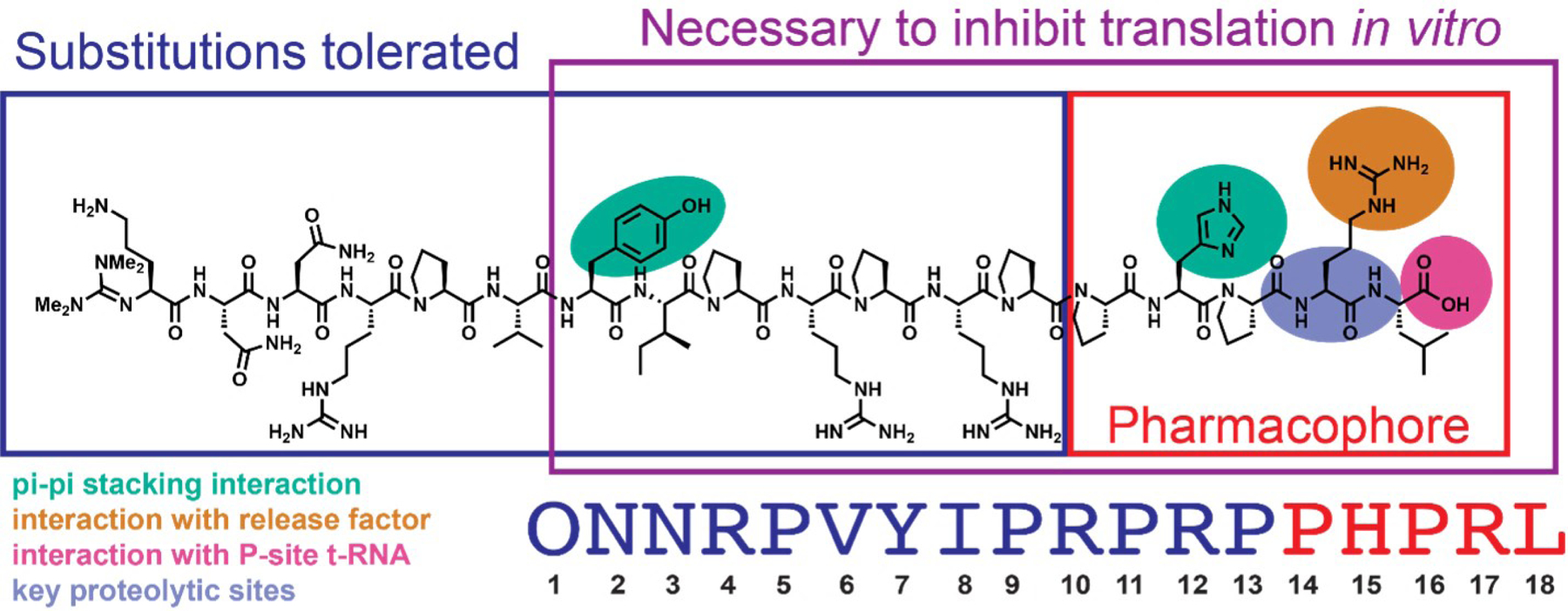
Figure 2.

Key residues in the sequence of Api-137 as per Baliga et al. The pharmacophore residues are boxed in red. The residues necessary to arrest the ribosome at the stop codon in vitro are boxed in purple. The residues which tolerate substitutions while retaining the activity of apidaecin endogenously expressed in E. coli cells are shown in navy blue. The Api137 residues of interest in this work: residues with π-π stacking interactions with rRNA of the nascent peptide exit tunnel of the 70S ribosome (turquoise), residues with interactions with the release factor (orange), residues with interactions with the P-site tRNA (magenta), and residues which are sites of proteolysis in murine serum40 (violet).
