
Figure 4. Interactions of Api-137 with the residues of the 23S rRNA.
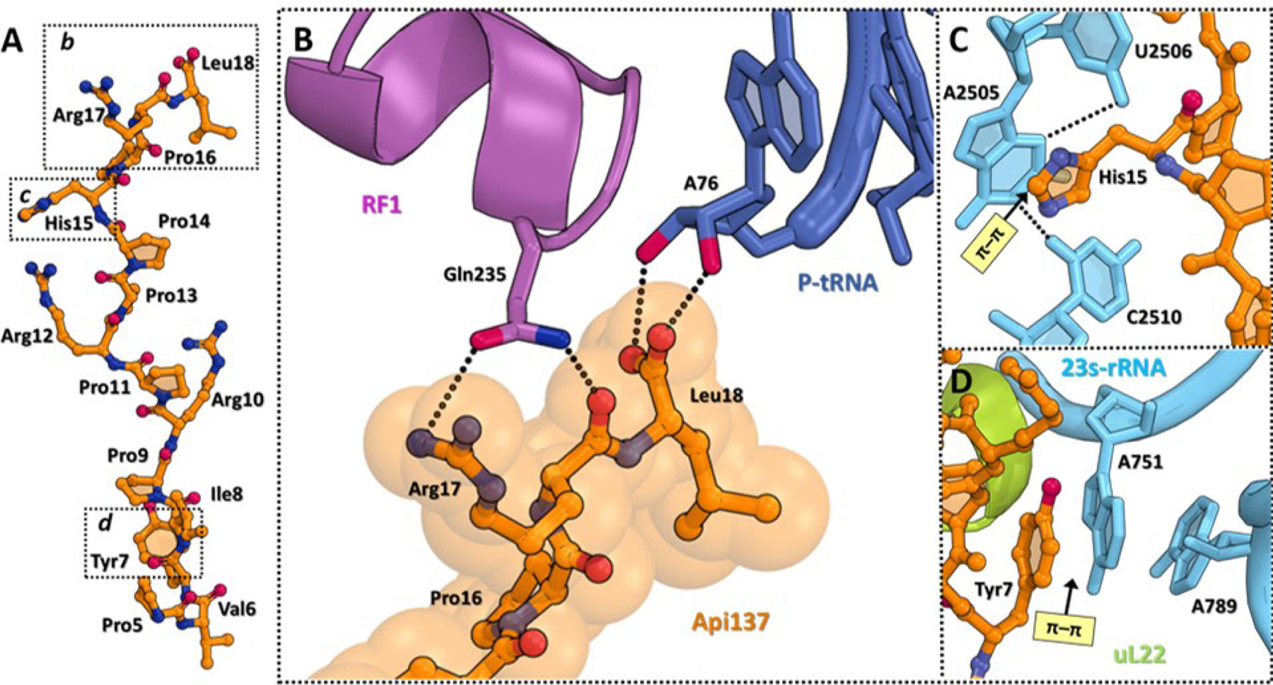
(A) Structure of ribosome-bound Api137 (orange). (B) Close-up view of the interactions of the C-terminal Arg17 and Leu18 residues of Api137 with the GGQ motif of RF1 (magenta) and A76 of the deacylated P-site tRNA (navy). H-bonds are shown with dotted lines. (C, D) π-π stacking interactions (light yellow) of Api residues (orange) with the nucleotides of the 23S rRNA (cyan). Ribosomal protein L22 is shown in light green. Figure rendered from PDB 5O2R.