
Figure 3.
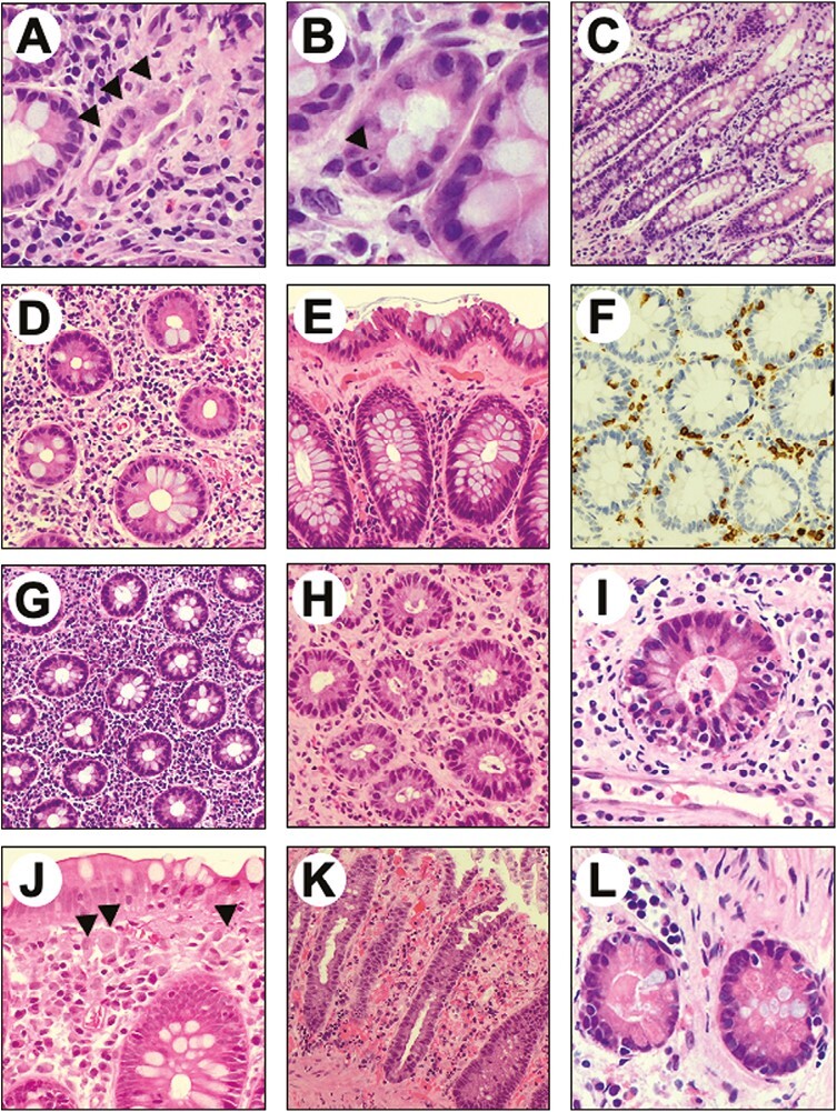
Representative examples of the microscopic alterations found in colic mucosa of patients treated with immune checkpoint inhibitors. (A) Crypt atrophy. (B) Apoptotic bodies within a gland. (C) Mild crypts’ distortion. (D) Lamina propria expansion and mucin depletion. (E) Collagenous band. (F). CD3 immunostaining showing moderate intraepithelial lympho-monocitic infiltrate. (G) Lympho-monocitic and plasmacellular infiltrate within the lamina propria. (H) Granulocytic infiltrate within the lamina propria associated with cryptitis. (I) Cryptic abscess. (J) Subepithelial macrophages. (K) Ischemic-like colitis features and superficial erosion. (L) Paneth metaplasia.