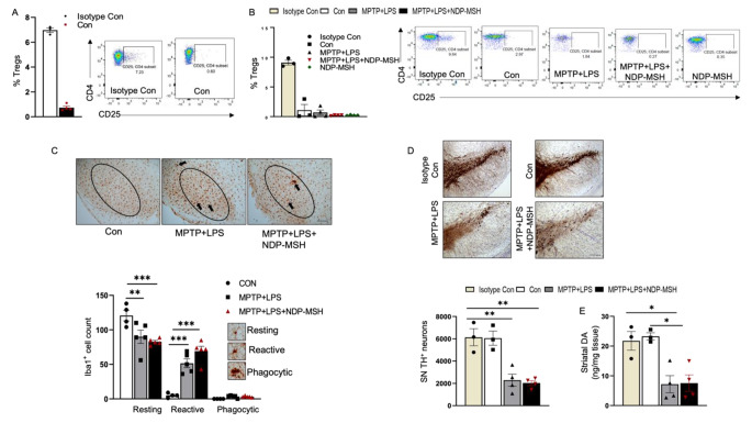
Fig. 4.
Depletion of Tregs abolishes neuroprotective effect of NDP-MSH. (A) C57BL/6 mice were pre-injected with PC61/CD25 antibody or isotype control. Mice were then injected with MPTP.HCl (20 mg/kg) + LPS (1 mg/kg) or vehicle (Con) and treated with NDP-MSH (400 µg/kg) or vehicle. Mice were sacrificed, and flow cytometry was carried out to assess the percentage of Treg cells in the spleen (A) before random grouping and (B) at the end of the treatment paradigm. (C) Representative micrograph staining for iba1 and morphological classification and quantification of iba1 + microglia in SN. scale bar 100 μm; n = 4/group. Two-way ANOVA followed by Tukey’s post hoc test; **p < 0.01; ***p < 0.001. (D) Representative micrograph of TH + staining and stereological quantification of TH + cells in SN. Scale bar, 100 μm; n = 3–4/group. One-way ANOVA followed by Tukey’s post hoc test. **p < 0.01. (E) Striatal dopamine content; n = 3–4/group. One-way ANOVA followed by Tukey’s post hoc test. *p < 0.05. Representative results from 2–3 independent experiments, which were conducted using preserved samples or fresh samples and were not pooled

