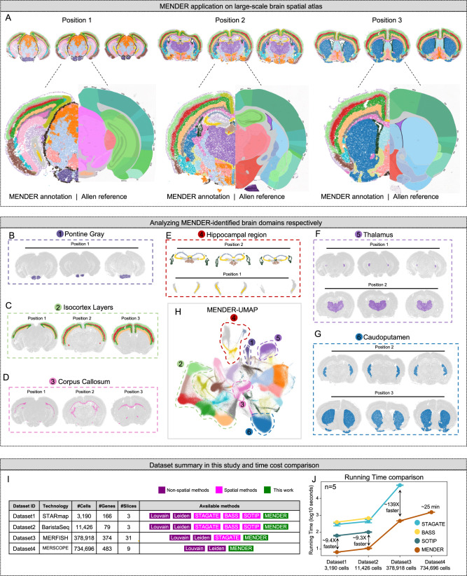
Fig. 4. Scalability and speed.
A–H Application of MENDER on a mouse brain atlas using MERSCOPE. A The dataset contains 9 slices from 3 different brain positions. For each slice, we show the MENDER result (top). We also highlight one slice for each position to compare with the Allen brain reference (bottom). Specific brain domains are shown, including Pontine Gray (B), Isocortex Layers (C), Corpus Callosum (D), Hippocampal region (E), Thalamus (F), and Caudoputamen (G). H The UMAP embedding on the top of MENDER cellular context representation. I Summary of benchmark datasets for unsupervised spatial domain identification. J Running time comparison of different spatial methods across the 4 datasets. Error bars are based on mean and 95% confidence interval. Source data are provided as a Source Data file.

