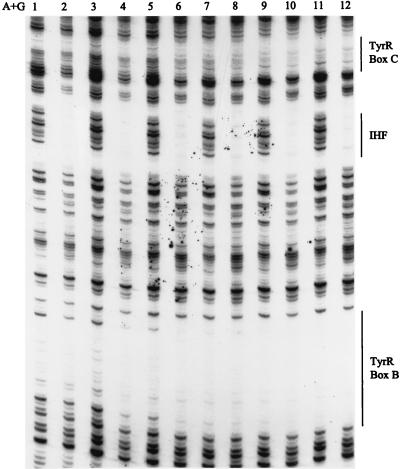
FIG. 7.
Effects of IHF and cAMP-CRP on DNase I footprinting of boxes B and C. The DNA used was 0.7 nmol of an EcoRI-PstI fragment from pUC19-tpl that had been 32P labeled at the 5′ end (see Materials and Methods). The tubes alone were used (lane 1), or CRP (20 nM) and IHF (145 nM) (lanes 2, 4, 6, 8, 10, and 12) were added to the tubes. The TyrR concentrations were as follows: lanes 2 and 3, 10 nM; lanes 4 and 5, 20 nM; lanes 6 and 7, 40 nM; lanes 8 and 9, 60 nM; lanes 10 and 11, 80 nM; lane 12, 100 nM. Each reaction mixture contained ATP and tyrosine (0.2 mM each). Tubes containing CRP also contained cAMP (100 μM).

