Fig. 1.
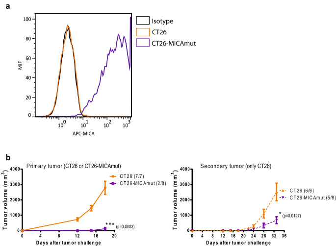
Stable expression of MICAmut by tumor cells promotes antitumor activity. (a) Determination of MICA expression by flow cytometry on CT26-MICAmut cell line. Orange and violet lines represent non-modified and MICAmut-lentiviral modified CT26 cell lines, respectively, and labelled with anti-MICA APC antibody. Black line indicates non-modified CT26 labeled with an isotype control. (b) left, tumor volume of balb/c mice injected subcutaneously with 1 × 10⁶ CT26 (orange) or CT26-MICAmut (violet) cells into the left flank; right, rechallenge performed 13 days after primary tumor implantation by injecting 1 × 10⁶ CT26 cells into the right flank of mice. The number of animals developing tumors out of the total number treated is indicated for each treatment. The mean of tumor volume ± SEM is shown. *p < 0.05, ***p < 0.001

