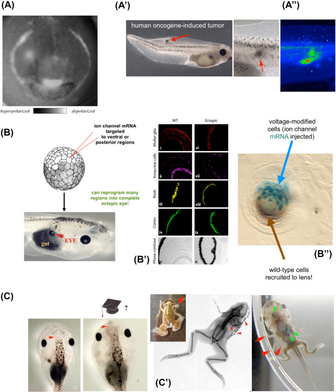
Fig. 5.
Examples of non-neural bioelectricity guiding behavior in morphospace. A Much as brain imaging allows the reading of bioelectrical states in tissues to decode the properties of control circuits and behavioral competencies, voltage reporter dyes enable in vivo tracking of the information processing in morphogenetic decision-making. Here is shown one frame from a timelapse video of a frog embryo prior to formation of the face, showing a prepattern of resting potential states that demarcates the position of the future gene expression domains and craniofacial organs, such as the eyes, mouth, and lateral structures. In contrast to this endogenous pattern, pathological patterns (such as those leading to tumors in A’) can be induced via, for example, oncogene injection. The location of tumors (A”, red arrowhead) can be predicted by the bioelectric dye signal which shows an aberrant electrical signature of cells that are disconnecting from the tissue-level network and reverting back to unicellular-scale behavior (i.e., metastasis and over-proliferation). B These bioelectric prepatterns are known to be instructive, because reproducing them elsewhere by misexpression of specific ion-channel mRNA, such as in this frog embryo, results in induction of whole organs, such as eyes (red arrow), which have the necessary internal tissue structure (immunohistochemistry in B’). This demonstrates a key aspect of behavior—binding complex downstream actions to a simple (low information-content) trigger. Moreover, this phenomenon exhibits the competency of recruitment (B”): when only a few cells are injected with the channel (cyan b-galactosidase marker label), they autonomously recruit normal neighbors to complete their morphological goal: building a normal-sized ectopic lens (brown tissue). C Method of inducing whole organs (control of large-scale movements in anatomical morphospace) by modulating bioelectric states of the tissue circuit (i.e., incepting false memories into the network) can also, for example, produce ectopic forebrain (red arrow), or ectopic limbs (C’, C”, red arrows). Panels reused with permission from (Chernet and Levin 2013a; Levin 2009; Levin et al. 2017; Pai et al. 2012; Vandenberg et al. 2011). (color figure online)

