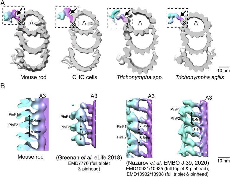
Figure S2. Comparison of pinhead architectures of complete triplets from different species.
(A) Cross-sectional views of microtubule triplets from mouse photoreceptor, CHO cells (Li et al, 2019), Trichonympha spp. (generic term for a group of termite gut protists), and Trichonympha agilis (Nazarov et al, 2020). All the pinheads (dashed box) connect to the A3 protofilament. (A, B) Longitudinal views of the pinheads (arrows in (A)). The pinheads have similar orientations, with the two pin feet moieties PinF1 and PinF2 extending from A3 PF. In T. spp, the average spacing between PinF1 and PinF2 of adjacent pinheads (7.9 nm) differs from that between PinF1 and PinF2 of the same pinhead (8.6 nm), whereas for the other species, these two spacings are the same: 8.4 nm for WT mouse, 8.2 nm for CHO, and 8.4 nm for T. agilis.

