FIGURE 1.

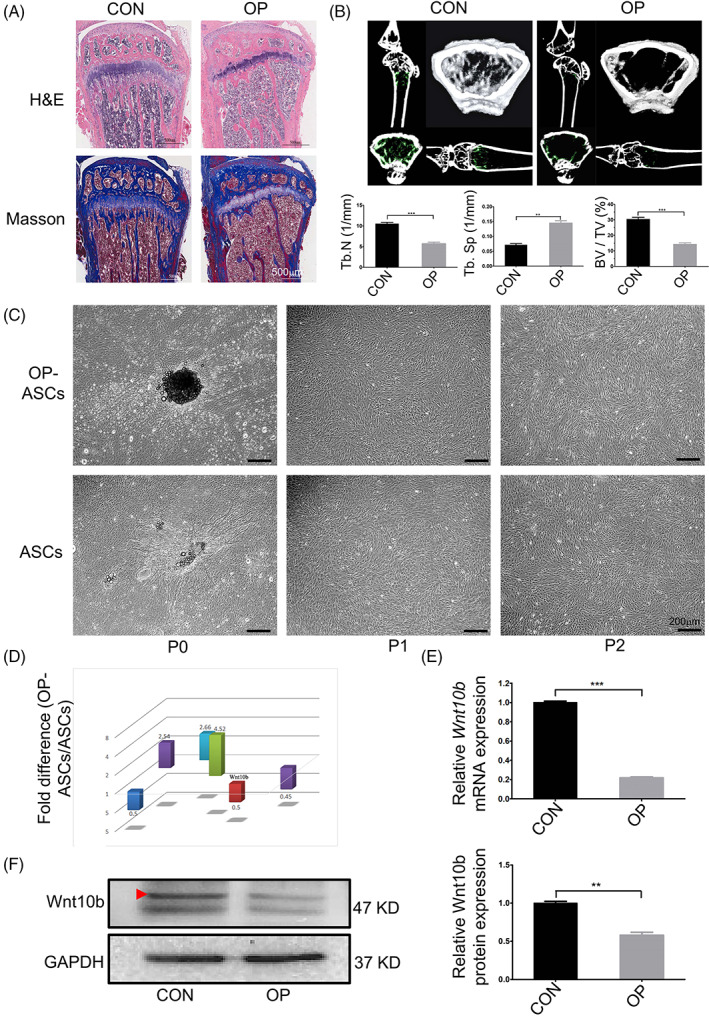
Wnt10b exhibited lower expression in OP‐ASCs than ASCs. (A) H&E and Masson's trichrome staining of the proximal metaphysis of the tibia from CON and OVX mice at 6‐week post‐operation. (B) The structure and morphology of the femur metaphysis from CON and OVX mice at 6‐week post‐operation obtained through micro‐CT, and statistical analysis of the Tb.N, BV/TV, and Tb.Sp between CON and OVX mice. (C) Images of OP‐ASCs and ASCs at different generations. (D) Expression of Wnt10b in OP‐ASCs was significantly lower than that observed in ASCs by GeneChip. (E) qPCR and statistical analysis showed that the mRNA expression level of Wnt10b was significantly decreased in OP‐ASCs when compared with ASCs. (F) Protein expression of Wnt10b in OP‐ASCs was lower than that in ASCs confirmed through WB. BV/TV, bone volume to tissue volume; CON, control; H&E, haematoxylin and eosin; micro‐CT, micro‐computed tomography; OP‐ASC, osteoporotic adipose‐derived stem cell; OVX, ovariectomized.
