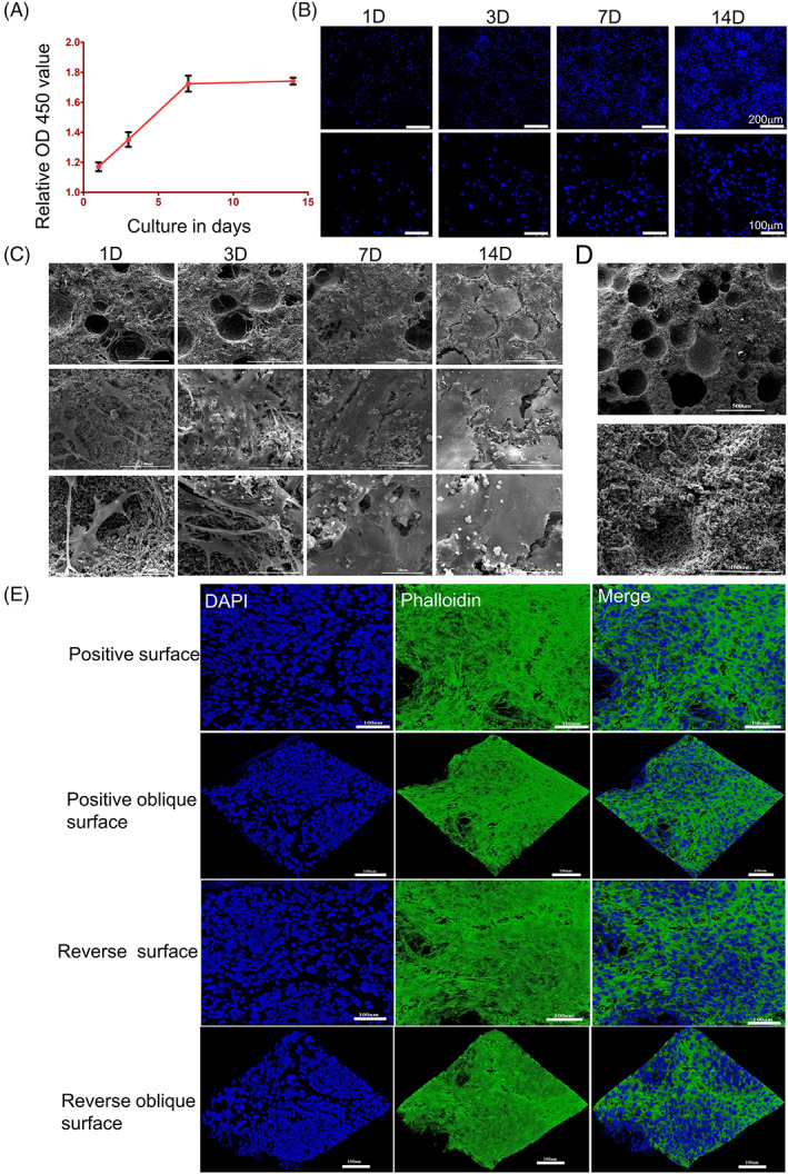
FIGURE 4.

OP‐ASCs could seed and proliferate well with BCP. (A, B) CCK‐8 and DAPI staining show the number of OP‐ASCs increased with time. The cell proliferation rate was faster at 1, 3 and 7 days of coculture, while at 7–14 days, the proliferation rate was significantly slower. (C) SEM scanning shows the same proliferation pattern as the above expression and confirms that OP‐ASCs combined well into the BCP scaffolds. (D) The physical properties of BCP. (E) Confocal laser scanning microscopy revealed OP‐ASCs were intertwined into a network and grew to saturation at 14 days of co‐cultivation. BCP, biphasic calcium phosphate; OP‐ASC, osteoporotic adipose‐derived stem cell; SEM, scanning electron microscope.
