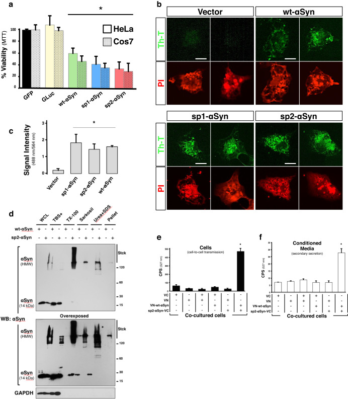
Fig. 2. sp2-αSyn is toxic, amyloidogenic and cell-to-cell transmitted.
a Determination of cell viability of HeLa and Cos7 cells expressing GFP, the luciferase of Gaussia princeps (GLuc), wt-, sp1- or sp2-αSyn by the MTT assay. Data is shown as the media ± SD. *p < 0.05 compared to GFP-expressing cells (one-way ANOVA followed by the post hoc Dunnett’s test, n = 5). b Representative confocal microscopy images of Cos7 cells expressing wt-, sp1- or sp2-αSyn and stained with the amyloid-specific dye thioflavin-T (Th-T). Cells transfected with an empty vector (Vector) were used as a negative control. Scale bar: 10 μm. c Quantification of Fig. 2b. *p < 0.05 (one-way ANOVA followed by the post hoc Tukey’s test, n = 5). d WCL of Cos7 cells expressing wt- or sp2-αSyn were subjected to sequential extraction by detergents and subsequently analyzed by WB. e, f HeLa cell clones expressing either VN or VC (N- and C- terminus half of the Venus fluorescent protein, respectively), or the fusion proteins VN-wt-αSyn or sp2-αSyn-VC were co-cultured as indicated. 48 h later the cells were harvested and fluorescence was quantified in living cells (e) or in the conditioned media to asses secondary secretion (f). Data is shown as the media ± SD. CPS counts per second. *p < 0.005 (one-way ANOVA followed by the post hoc Tukey’s test, n = 5).

