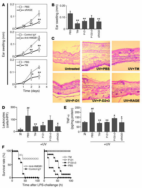
Figure 3.
Central role of HMGB1 in UV irradiation–induced skin injury and the effect of TM and TM-derived peptides on HMGB1-mediated local and systemic inflammation. (A) In vivo effect of HMGB1 blockade on UV irradiation–induced cutaneous inflammation. The time course for ear swelling was measured after local subcutaneous administration of PBS or sRAGE (3 pmol; top); anti-HMGB1 IgY (Anti-HMGB1) or nonimmune IgY (Control IgY, 300 ng; middle); or rhs-TM (3 pmol) or PBS before exposure to UV irradiation (bottom). In each case, data shown are mean ± SD (n = 8). *P < 0.05; **P < 0.01. (B–E) As indicated, mice received PBS, rhs-TM, P-D1, P-D2+3 (each at 100 nmol/kg; i.p.) or sRAGE (25 nmol/kg; i.p.) at 1 hour and 12 hours after exposure to UV irradiation (n = 8 per group). Three days after UV treatment, ear swelling (B), general histopathological features of the skin (C; H&E staining; magnification, ×100), infiltrating leukocytes (D; cells/HPF), and local TNF-α accumulation (E) were assessed. The mean ± SD of 6 determinations is shown (B, D, and E). *P < 0.05 and **P < 0.01, compared with the UV-irradiated control group given PBS. ##P < 0.01, compared with untreated controls. (F) Effect of TM and TM-derived peptides on systemic LPS-induced lethal challenge. Left panel: mice (BALB/c) received LPS (10 mg/kg; i.p.) in the presence of anti-HMGB1 IgY or nonimmune IgY (3 i.p. doses of 2 mg/kg/mouse of IgY at 2, 12, and 24 hours after LPS). Right panel: mice were treated with LPS as described above and also received rhs-TM, P-D1, P-D2+3, or PBS (3 i.p. doses of 100 nmol/kg/mouse for each peptide).

