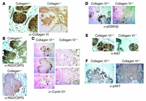
Figure 4.
Immunohistochemical analysis of pathologically matched tumors from collagen+/+ and collagen–/– mice in the MMTV-PyMT background. (A) Absence of collagen VIα3 signal in collagen–/– mice. Staining for collagen VIα3 (with carboxyterminal polyclonal antibody) on mammary sections from pathologically matched hyperplasias developed in collagen+/+ and collagen–/– mice. Arrows point in the direction of increased levels of staining. Asterisks denote local mammary adipocytes. Magnification, ×25. (B) NG2/CSPG is expressed at the highest levels in malignant regions adjacent to adipocytes in WT mice. Note the positive staining in WT backgrounds on the leading edges of the tumor mass. Magnification, ×25. (C) Cyclin D1 is expressed at the highest levels in malignant regions adjacent to adipocytes. Magnification, ×10. (D) GSK3β is phosphorylated at the highest levels in malignant regions adjacent to adipocytes. Magnification, ×10. (E) Ki67 expression is present in cells proliferating throughout the mammary lesions. Staining for the proliferation marker Ki67 in pathologically matched lesions from WT and collagen VI–null mice demonstrates diffuse expression throughout the tumor masses in both genotypes. Magnification, ×25. (F) Akt is phosphorylated and activated at higher levels in tumors that have developed in the presence of collagen VI signaling. Staining for the ser473-phosphorylated Akt in pathologically matched tumors from collagen VI WT and knockout mice demonstrated significant staining in cells on the tumor periphery in the WT mice but not in collagen VI–null mice. Asterisks denote local mammary adipocytes in close proximity to the malignancies. Arrowheads point to the pAkt-positive cells. Magnification, ×25.

