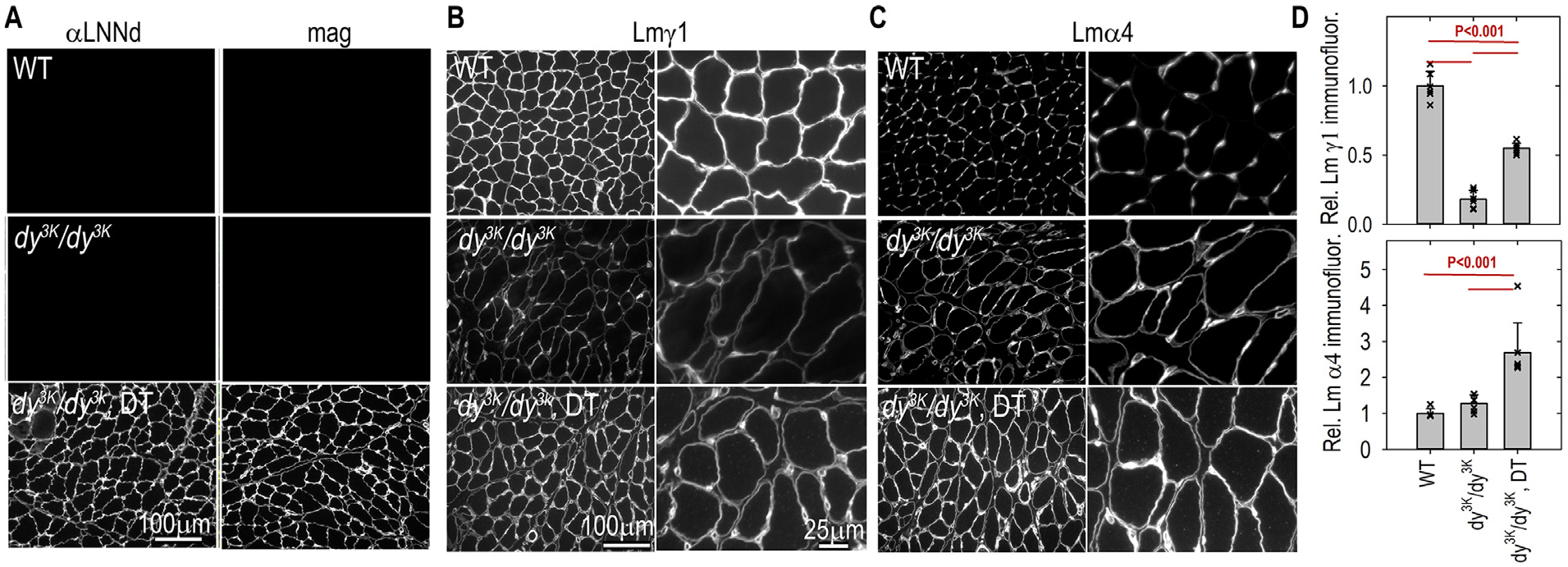
Fig. 8. Laminin muscle immunofluorescence.

Frozen sections of hindlimb muscle from 3-week-old mice were immunostained with antibodies to detect linker proteins and laminin subunits. Panel A: antibodies to Lmα1 LN-LEa domains (αLNNd) and mag. Panel B: laminin-γ1. Panel C: laminin α4. Panel D: Morphometry comparing the sum of BM intensities (av. ± s.d. with individual image values (symbols) shown. Both subunits were reduced in dy3K/dy3K muscle. DT-dy3K/dy3K mice revealed an increase in Lmγ1 to levels about half-way between WT and dy3K/dy3K without transgenes whereas Lmα4 levels were significantly higher in DT-dy3K/dy3K compared to WT and dy3K/dy3K mice.
