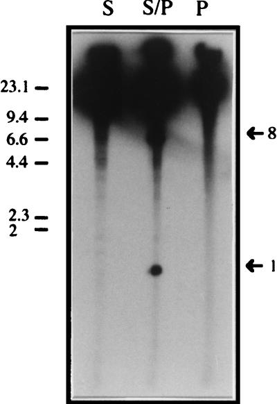
FIG. 2.
Identification of genomic fragments of P. putida KT2440 smaller than 15 kb after digestion with SwaI, PmeI, and SwaI plus PmeI and 32P end labeling. Total DNA of P. putida KT2440 genomic DNA was digested with SwaI (lane S), PmeI (lane P), and SwaI plus PmeI (lane S/P) and then end labeled with 32P as described in Materials and Methods. Fragments were separated in a conventional 0.8% (wt/vol) agarose gel run in 1× TAE buffer for 3 h at 5 V cm−1. The gel was exposed to Kodak photographic film and developed. Size fragments are indicated in kilobases. Bands of interest and their sizes are indicated by arrows.

