

Summary
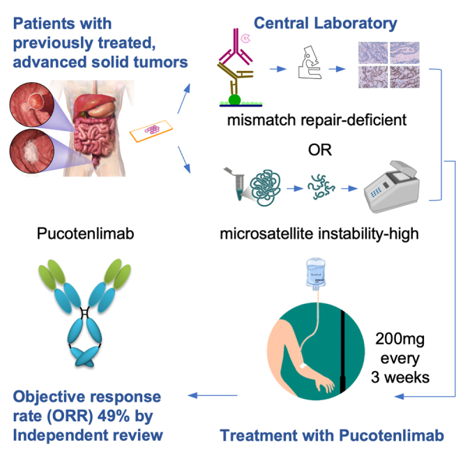
We report a multicenter, phase 2 study evaluating the efficacy of pucotenlimab, an anti-PD-1 antibody, in patients with mismatch repair-deficient (dMMR) or microsatellite instability-high (MSI-H) tumors, and potential biomarkers for response. Overall, 100 patients with previously treated, advanced solid tumors centrally confirmed as dMMR or MSI-H received pucotenlimab at 200 mg every 3 weeks. The most common cancer type is colorectal cancer (n = 71). With a median follow-up of 22.5 months, the objective response rate is 49.0% (95% confidence interval 38.86%–59.20%) as assessed by the independent review committee, while the median progression-free survival and overall survival have not been reached. Grade ≥3 treatment-related adverse events were observed in 18 patients. For the biomarker analysis, responders are enriched in patients with mutations in the KMT2D gene. Pucotenlimab is an effective treatment option for previously treated advanced dMMR/MSI-H solid tumors, and the predictive value of KMT2D mutation warrants further research. This study is registered with ClinicalTrials.gov: NCT03704246.
Keywords: mismatch repair deficient, microsatellite instability-high, solid tumor, efficacy, safety, biomarker
Graphical abstract
Highlights
-
•
Pucotenlimab is a humanized anti-PD-1 monoclonal antibody with an engineered Fc domain
-
•
Patients with advanced dMMR/MSI-H tumors derive durable responses to pucotenlimab
-
•
Treatment-related adverse events are manageable
-
•
KMT2D mutation warrants further investigation as a predictive biomarker
Zhang et al. report the outcomes of a phase 2 study evaluating pucotenlimab, an anti-PD-1 antibody, in patients with previously treated, advanced mismatch repair-deficient or microsatellite instability-high solid tumors. Pucotenlimab demonstrates durable anti-tumor activity with manageable toxicities. Additionally, KMT2D might be a potential biomarker for response in this patient population.
Introduction
The incidence rate of mismatch repair-deficient (dMMR) or microsatellite instability-high (MSI-H) tumors was approximately 2%–4% of all diagnosed cancers.1,2 The frequency of dMMR/MSI-H is higher in several cancer types, including endometrial cancers, gastric cancers, and colorectal cancers.3 The mismatch repair genes include MLH1, MSH2, MSH6, and PMS2, and the status of dMMR or MSI-H is evaluated at the protein and DNA level, respectively. Tumors with dMMR/MSI-H exhibit 10 to 100 times more mutations than mismatch repair-proficient or microsatellite stable tumors and lead to a high tumor mutational burden, enhancing expression of neoantigens and increasing lymphocytes infiltration.4,5 These immunogenic features provide a strong rationale for immune checkpoint inhibition in patients with dMMR/MSI-H tumors.
Anti-PD-1 antibodies have demonstrated promising activity in several clinical studies involving patients with dMMR/MSI-H tumors. Among them, pembrolizumab was approved by the Food and Drug Administration of the United States for the first, second, or later line treatment of metastatic dMMR/MSI-H colorectal cancer (CRC),6,7 nivolumab was approved for metastatic dMMR/MSI-H CRC previously treated with a fluoropyrimidine, oxaliplatin, and irinotecan,8 and dostarlimab was approved for previously treated dMMR/MSI-H solid tumors.9 However, one of the major challenges in the management of patients with advanced dMMR/MSI-H tumors is that only 30%–40% of patients respond to PD-1 blockade, whereas around 30% patients have intrinsic resistance to the treatment.6,7,8,9,10 Therefore, there remains a critical need for reliable predictive biomarkers in these patients for durable clinical response.
Pucotenlimab is a humanized IgG4S228P anti-PD-1 monoclonal antibody with an engineered Fc domain.11 Earlier studies have demonstrated the potential anti-tumor activity and manageable safety profile of pucotenlimab in the patients with solid tumors.12,13 We conducted a prospective phase II study with two parallel cohorts to define the efficacy and safety of pucotenlimab in patients with advanced gastric or gastroesophageal junction cancer (cohort 1) and advanced dMMR/MSI-H solid tumors (cohort 2), respectively; the findings from cohort 1 have been reported.14 Here, we present the clinical outcomes and results of biomarker analyses from cohort 2.
Results
Patient characteristics
From October 25, 2018, to December 3, 2020, we screened 1,392 patients for eligibility, and finally a total of 100 patients with centrally determined dMMR/MSI-H locally advanced or metastatic solid tumors were recruited, comprising the intention-to-treat (ITT) population. At data cutoff (December 4, 2021), the median follow-up was 22.5 (range: 0.3–37.4) months. The median age was 53 years (range: 20–74), and 54 of them were male. The most common cancer types were CRC (n = 71), gastric cancer (n = 10), and endometrial cancer (n = 7). Ninety-eight patients had distant metastatic tumors, while the other two had locally advanced disease. All patients had received previous systemic therapies for advanced disease, and 44 of them had received at least two prior lines of treatment. Ninety-seven patients were enrolled with disease progression on previous systemic treatment, while three patients were enrolled with chemotherapy intolerance. Among the 71 patients with CRC, 30 had received prior therapies including a fluoropyrimidine, oxaliplatin, and irinotecan, 25 had received bevacizumab, seven had received cetuximab, while six had received multi-kinase inhibitors that target vascular endothelial growth factor receptors. All 10 patients with gastric cancer had HER2-negative tumors. The baseline characteristics of the patients are summarized in Table 1.
Table 1.
Patient characteristics at baseline (n = 100)
| Characteristics | n (%) | |
|---|---|---|
| Age | median age, years (range) | 53 (20–74) |
| age ≥65 years | 18 (18.0) | |
| Sex assigned at birth | male | 54 (54.0) |
| ECOG performance status | 0 | 21 (21.0) |
| 1 | 79 (79.0) | |
| Cancer type of primary diagnosis | colorectal cancer | 71 (71.0) |
| gastric cancer | 10 (10.0) | |
| endometrial cancer | 7 (7.0) | |
| neuroendocrine tumor | 4 (4.0) | |
| ovarian cancer | 3 (3.0) | |
| cervical cancer | 2 (2.0) | |
| esophageal cancer | 2 (2.0) | |
| anal cancer | 1 (1.0) | |
| Centrally assessed MMR/microsatellite status | dMMR and MSI-H | 81 (81.0) |
| dMMR only | 12 (12.0) | |
| MSI-H only | 7 (7.0) | |
| Lines of prior systemic therapies | 1 | 56 (56.0) |
| ≥2 | 44 (44.0) | |
| Lines of prior systemic therapies for patients with colorectal cancer (n = 71) | 1 | 43 (43.0) |
| ≥2 | 28 (28.0) | |
| Disease stage | locally advanced | 2 (2.0) |
| distant metastatic | 98 (98.0) | |
| Metastatic sites | liver metastasis | 41 (41.0) |
| adrenal gland metastasis | 2 (2.0) | |
| lung metastasis | 12 (12.0) | |
| bone metastasis | 10 (10.0) | |
| pleural effusion | 7 (7.0) | |
| peritoneal effusion | 11 (11.0) | |
| Lynch syndrome as confirmed by germline sequencing | yes | 27 (27.0) |
| no | 57 (57.0) | |
| not performed | 16 (16.0) | |
See also Table S1 and Table S3.
Abbreviations: ECOG, Eastern Cooperative Oncology Group.
At data cutoff, 48 of 100 patients were still on treatment. The most common reasons for treatment discontinuation were disease progression (n = 35) and adverse events (n = 12). The median dose number of pucotenlimab received was 20 (ranged 1–53). The study flowchart is shown in a CONSORT diagram (Figure S1).
Efficacy
Among the 100 patients enrolled, 93 patients were evaluable for tumor response, and 7 patients were not evaluated. Based on the assessment by the independent review committee (IRC), nine patients achieved confirmed complete response (CR), 40 achieved confirmed partial response (PR), 15 had stable disease (SD), while six patients had non-CR/non-PD (N/N), thus the objective response rate (ORR) was 49.0% (95% confidence interval [CI] 38.86%–59.20%) and the disease control rate (DCR) was 70.0% (95% CI 60.02%–78.76%). Both the median progression-free survival (PFS) and median duration of response (DOR) were not reached, and the 12-month PFS rate was 56.1% (95% CI 45.63%–65.24%). The median overall survival (OS) was not reached, and the 24-month OS rate was 68.9% (95% CI 57.99%–77.45%). Similarly, upon investigators assessment, the ORR was 50.0% (95% CI 39.83%–60.17%) and DCR was 73.0% (95% CI 63.20%–81.39%); the median PFS was 22.8 (6.18, NR) months, and the 12-month PFS rate was 52.7% (95% CI 42.45%–61.96%). The efficacy outcomes are shown in Table 2 and Figure 1.
Table 2.
Efficacy per investigator and independent review committee’s assessment for all patients and patients with colorectal cancer
| All patients (n = 100) | Patients with CRC (n = 71) | ||
|---|---|---|---|
| IRC | Investigator | IRC | |
| CR, n (%) | 9 (9.0) | 9 (9.0) | 6 (8.5) |
| PR, n (%) | 40 (40.0) | 41 (41.0) | 34 (47.9) |
| Non-CR/Non-PD, n (%) | 6 (6.0) | – | 2 (2.8) |
| SD, n (%) | 15 (15.0) | 23 (23.0) | 8 (11.3) |
| PD, n (%) | 23 (23.0) | 20 (20.0) | 15 (21.1) |
| NE, n (%) | 7 (7.0) | 7 (7.0) | 6 (8.5) |
| ORR, % (95% CI) | 49.0 (38.86, 59.20) | 50.0 (39.83, 60.17) | 56.3 (44.05, 68.09) |
| DCR, % (95% CI) | 70.0 (60.02, 78.76) | 73.0 (63.20, 81.39) | 70.4(58.41, 80.67) |
| Median PFS, months (95% CI) | NR | NR | NR |
| 12-month PFS rate, % (95% CI) | 56.1 (45.63, 65.24) | 52.7 (42.45, 61.96) | 58.2(45.65, 68.77) |
| Median DOR, months (95% CI) | NR | NR | NR |
| Median OS, months (95% CI) | NR | NR | |
| 24-month OS rate, % (95% CI) | 68.9 (57.99, 77.45) | 73.5(60.33, 82.96) | |
Abbreviations: CRC, colorectal cancer; IRC, independent review committee; CR, complete response; PR, partial response; SD, stable disease; PD, progressive disease; NE, not evaluated; ORR, objective response rate; DCR, disease control rate; PFS, progression-free survival; CI, confidence interval; DOR, duration of response; OS, overall survival; NR, not reached.
Figure 1.

Tumor response in patients with previously treated dMMR/MSI-H solid tumors per RECIST v1.1 based on IRC’s assessment
(A) Percentage changes from baseline in the sum of the tumor burden for target lesions over time until first progressive disease (if any).
(B) The best percentage changes from baseline in the sum of the tumor burden for target lesions before the first progressive disease (if any). The results of the KMT2D mutational status are also shown in the plot.
(C) The swimmer plot showing the key events during the treatment for each patient.
The dotted lines in (A) and (B) indicate a 30% reduction or 20% increase.
Specifically, among the 71 patients with advanced CRC, the ORR was 56.3% and the DCR was 70.4% according to IRC’s assessment. With a median follow-up of 23.5 months, the median PFS was not reached, and the 12-month PFS rate was 58.2%. The efficacy outcomes of patients with advanced CRC are shown in Table 2.
Safety
All the 100 enrolled patients had received at least one dose of pucotenlimab and were thus included in the safety analysis. Overall, 83 patients (83%) had treatment-related adverse events (TRAEs) and 18 (18%) had grade 3 and above TRAEs. The only fatal event was one case of grade 5 myocarditis. Five patients (5%) had treatment-related serious adverse events (SAEs), and four (4%) discontinued treatment because of a TRAE. The most common TRAEs of any grade were aspartate aminotransferase (AST) increased (n = 26; 26.0%), alanine aminotransferase (ALT) increased (n = 23; 23.0%), anemia (n = 22; 22.0%), and hypothyroidism (n = 20; 20.0%). The most frequent grade 3 TRAEs were anemia (4.0%), leukopenia (2.0%), and hypertension (2.0%), while only one case of grade 4 treatment-related adverse event was observed (neutropenia).
Immune-mediated adverse events (irAEs) and infusion reactions, regardless of attribution to treatment by the investigators, were observed in 29 (29%) patients, of which three (3%) were immune-related SAEs. Three patients (3%) discontinued treatment because of an irAE or infusion reaction. The most common irAEs were hypothyroidism (12.0%), hyperthyroidism (5.0%), and thyroid function test abnormal (3.0%). Most events were grade 1 and 2 in severity. Grade 3 irAEs included type 1 diabetes mellitus in one patient, and one case of pneumonitis, myositis, and myocarditis at the same time. Grade 5 irAE was observed in one patient who died of multiple factors including immune-mediated myocarditis and disease progression. TRAEs and irAEs are summarized in Table 3.
Table 3.
Safety profile for all the patients enrolled (n = 100)
| Adverse eventa | Any grade, n (%) | Grade ≥3b, n (%) |
|---|---|---|
| TRAEs | 83 (83) | 18 (18) |
| Occurring in ≥10% of patients | ||
| AST increased | 26 (26) | 0 |
| ALT increased | 23 (23) | 0 |
| Anemia | 22 (22) | 4 (4) |
| Hypothyroidism | 20 (20) | 0 |
| Blood bilirubin increased | 19 (19) | 0 |
| Thyroid function test abnormal | 16 (16) | 0 |
| Leukocyte count decreased | 16 (16) | 2 (2) |
| Neutrophil count decreased | 12 (12) | 1 (1) |
| Rash | 12 (12) | 0 |
| γ-GT increased | 11 (11) | 0 |
| Platelet count decreased | 10 (10) | 0 |
| irAEsc reported in >1% of patients | ||
| Hypothyroidism | 12 (12) | 0 |
| Hyperthyroidism | 5 (5) | 0 |
| Thyroid function test abnormal | 3 (3) | 0 |
| ALT increased | 2 (2) | 0 |
| AST increased | 2 (2) | 0 |
| Blood bilirubin increased | 2 (2) | 0 |
| Myositis | 2 (2) | 1 (1) |
Abbreviations: TRAE, treatment-related adverse event; AST, aspartate aminotransferase; ALT, alanine aminotransferase; irAE, immune-related adverse event.
Events are listed in order of descending frequency. Safety analyses included all patients who received at least one dose of pucotenlimab.
There was one case of grade 4 immune-mediated adverse event (type 1 diabetes mellitus) and one case of grade 5 treatment-related adverse event (myocarditis).
Based on a list specified by the sponsor and considered regardless of attribution to treatment or immune relatedness by investigator.
Biomarker analysis
PD-L1 expression was performed in 79 patients, whereas 21 patients did not provide adequate samples for testing. The tumor samples of 46 patients had PD-L1 tumor proportion score (TPS) ≥1%, 33 had PD-L1 TPS <1%. Patients with PD-L1 TPS ≥1% and TPS <1% had similar ORRs (54.3% vs. 48.5%, p = 0.775).
Neutrophil-to-lymphocyte ratio (NLR) was analyzed in 87 patients (Table S1). According to baseline NLR, 56 patients had NLR <4 and 31 patients had NLR ≥4, and there was no statistical difference in ORR and PFS between the two groups (ORR 60.7% vs. 48.4%, p = 0.267, median PFS not reached [NR] vs. NR, p = 0.302). Patients were then separated into two groups according to NLR at the second treatment cycle of pucotenlimab. There were 65 patients with NLR <4 and 21 patients with NLR ≥4 (one patient discontinued treatment due to irAEs). The ORRs of patients with NLR <4 and ≥4 at the second treatment cycle were 66.2% and 23.8% (p = 0.001), respectively. Patients with NLR <4 at the second treatment cycle also showed improved PFS and OS compared with those with NLR ≥4 (Figure S2). Overall, NLR decreased after treatment with pucotenlimab, which was contributed by both decrease in neutrophil count and increase in lymphocyte count in all patients included in the analysis. When the dynamics of NLR, neutrophil, and lymphocyte counts were analyzed with the patients’ best response to pucotenlimab, only patients who responded had marked on-treatment decrease in NLR (Figure S3), decrease in neutrophil count, and increase in lymphocyte count. Further, when the subgroups of lymphocytes were examined by flow cytometry, the increase in lymphocyte count was contributed by an increase in CD16/56+ lymphocytes, instead of alterations in CD3+, CD4+, or CD8+ lymphocyte counts.
Germline human leukocyte antigen class I (HLA-I) zygosity was determined in 84 patients. Among them, 71 patients (84.5%) were heterozygous in all HLA-I loci, and 13 patients (15.5%) were homozygous at one HLA-I locus. In the 73 patients who were analyzed for loss of heterozygosity (LOH) of HLA-I genes in their tumor samples, 38 had LOH of HLA-I. We observed similar ORRs in patients with homozygous or heterozygous germline HLA-I (46.2% vs. 53.5%, p = 0.852) and in patients with or without LOH of HLA-I (47.4% vs. 62.9%, p = 0.274).
Tumor samples from 73 patients were collected to assess tumor mutational burden (TMB) and genetic alterations in the tumor. The most common cancer types included CRC (n = 50), gastric cancer (n = 9), and endometrial carcinoma (n = 7).
The median TMB of the 73 patients was 41.30 mut/Mb (range: 0.87–229.57 mut/Mb). The median TMB in patients having objective responses (n = 40) was 42.39 mut/Mb (range: 0.87–108.26 mut/Mb), whereas the median TMB in non-responders (n = 33) was 29.13 mut/Mb (range: 1.30–229.57 mut/Mb). The cutoff point to distinguish TMB-low and TMB-high tumors was 32.5 mut/Mb, corresponding to the lower 35th percentile in this cohort. Patients with TMB-high tumor (n = 48) had a higher ORR compared with patients with TMB-low tumor (n = 25) (68.7% vs. 28.0%, p = 0.002).
In the analysis of the association of genetic alterations with clinical response, a cutoff of 0.05 was used for variant allele frequency, filtering out those low-frequency variants that may not be biologically relevant. The most commonly mutated genes revealed by next-generation sequencing (NGS) were KMT2D, TGFBR2, ARID1A, and KMT2B. In our screen for potential genetic alterations that were associated with clinical efficacy using the random forest model, the sliding windows sequential forward feature selection algorithm identified the top 21 important genes. The process of gene selection is presented in the supplemental information (Figure S4). Among the 21 genes that were subsequently evaluated with logistic regression, we observed that only patients harboring mutated KMT2D (n = 53) had significantly higher ORR (69.8% vs. 15%; adjusted p = 0.009 [using the Bonferroni method]) than those with wild-type KMT2D (n = 20), as shown in the supplemental information (Table S2). We further adjusted the result taking TMB as a potential confounding factor, and the difference remained significant (adjusted odds ratio: 6.24 [95% CI, 1.6–24.38], p = 0.008). No significant difference in ORR was observed according to mutation status in KRAS, NRAS, HRAS, and BRAF genes, which are commonly tested in patients with metastatic CRC. Specifically, among the 25 patients with KRAS mutations, the most common forms of mutation were KRASG12D (n = 10) and KRASG13D (n = 10), while the ORRs in patients having these two mutations were 50.0% and 80.0%, respectively.
In our exploratory multivariate regression analyses, NLR <4 at the second treatment cycle and KMT2D mutation were associated with improved ORR. According to the multiple Cox regression analyses, NLR <4 at the second treatment cycle, and KMT2D mutation were associated with longer PFS, whereas NLR <4 at the second treatment cycle, and TMB-high (TMB ≥ 32.5 mut/Mb) were associated with longer OS (Table S3).
Discussion
In the current study, pucotenlimab showed durable anti-tumor activity and manageable toxicities for the treatment of previously treated advanced dMMR/MSI-H solid tumors. Thus, our results support the use of pucotenlimab in this patient population. More importantly, we performed a comprehensive biomarker analysis to uncover potential predictors of efficacy. The findings from our present study showed that KMT2D mutations were associated with improved response to PD-1 blockade in patients with dMMR/MSI-H tumors, and thus they hold promise as a predictive biomarker for this patient population. We also identified that NLR after treatment with an PD-1 inhibitor in patients with dMMR/MSI-H tumors correlated with response, and that both decrease in neutrophil count and increase in CD15/56+ lymphocytes contributed to the dynamics of NLR in responders. Moreover, among biomarker studies in patients with dMMR/MSI-H tumors, our analyses involved the largest sample size from a prospective study, and the prospective nature ensured high-quality data regarding patients’ outcomes.
The ORR based on IRC’s assessment reached 49% in the 100 patients enrolled in our study, and the 12-month PFS and OS rates were 56.1% and 68.9%, respectively. Specifically, the ORR among patients with metastatic CRC, the most common cancer type in our cohort, reached 56.3%. Notably, responses to pucotenlimab were durable, since the median PFS was still immature when the median follow-up was approaching 2 years. In a previous meta-analysis evaluating the efficacy of immune checkpoint inhibitors in patients with MSI-H advanced malignancies, mainly in the pretreated setting, the pooled ORR was 41.5%, and the ORR for the subgroup of patients with CRC was 47.1%.15 According to the results of another meta-analysis, the ORR with PD-1/PD-L1 inhibitors in patients with dMMR/MSI-H advanced CRC was 37%.16 Although the results may not be directly comparable, the historical context highlighted the promising efficacy observed in our current study.
The safety profile of pucotenlimab, including the incidence of grade 3 or above TRAEs, SAEs, and fatal TRAEs, was consistent with those previously reported and with those of other PD-1 inhibitors.6,7,8,9,10,12,13,14 The pattern of immune-mediated adverse events and infusion reactions was as anticipated. Overall, pucotenlimab was well tolerated.
In our study, ORR did not differ between patients with PD-L1-positive (TPS ≥ 1%) and PD-L1-negative (TPS < 1%) tumors, although PD-L1 expression is frequently used in patient selection for the treatment with immune checkpoint inhibitors in several cancer types. Similar findings have also been observed in the CheckMate-142 study, in which nivolumab was given to patients with previously treated dMMR/MSI-H CRC, and those with PD-L1 ≥1% achieved an ORR of 29%, while those with PD-L1 <1% achieved an ORR of 28%.8 Hence, PD-L1 expression seems to lose its value as a predictor for efficacy in the context of dMMR/MSI-H tumors.
Patients with NLR below a cutoff of 4 at the second cycle of pucotenlimab treatment demonstrated greater chance of responding to the treatment than those with NLR above 4 in our study. The underlying mechanisms for the predictive value of NLR included that higher level of lymphocytes facilitate the host immune response to malignancy,17 and that inflammation manifested by the increased peripheral neutrophil count plays an important role in various pathological processes involving tumor growth.18 Indeed, previous studies have reported that lower NLRs of either baseline or post initial treatment cycles were associated with better outcomes with PD-1 blockade in several tumor types.18,19 The ORRs observed in our study suggest that NLR could be an easy-to-access tool to predict outcomes in patients with dMMR/MSI-H tumors. We further discovered that pucotenlimab induced the decrease of neutrophil count and the increase of CD16/56+ natural killer (NK) cell counts in responders. NK cells have been found to mediate the effect of PD-1/PD-L1 blockade in addition to cytotoxic T cells in cancer.20 In patients with non-small cell lung cancer, a significant increase of NK cell count after the treatment of anti-PD-1 antibodies was observed in responders but not in non-responders in a previous study.21 It remains to be investigated if the increase in peripheral NK cell counts is associated with NK cell-dependent anti-cancer immunity in the tumor microenvironment induced by PD-1 inhibition.
It has been suggested that germline homozygosity in at least one HLA-I locus could lead to presentation of a smaller and less diverse repertoire of tumor-derived neoantigens to cytotoxic T lymphocytes. Meanwhile, HLA LOH with loss of either maternal or paternal allele of HLA in the tumor is one of the key mechanisms of immune disruption and tumor invasion.22,23 These mechanisms might affect patients’ outcomes when treated with immunotherapy. The hypothesis has been proved in a previous work that demonstrated a significant survival advantage to HLA-I heterozygosity both at the germline and somatic level in patients treated with immune checkpoint inhibitors.24 In our study, germline and somatic HLA-I heterozygosity were not associated with superior response to pucotenlimab. A possible explanation could be that the higher TMB in patients with dMMR/MSI tumors partly overcame the potential deficiency in antigen presentation. Therefore, a combination with TMB could be useful to improve the value of HLA-I heterozygosity as a biomarker in patients with dMMR/MSI tumors treated with PD-1 inhibitors, while the association of HLA-I zygosity with survival still awaits longer follow-up duration.
Consistent with previous reports, a high TMB is predictive of response to pucotenlimab in patients dMMR/MSI-H cancers.25,26 However, no validated cutoff values have been established for patient selection in this population, therefore limiting its use. The NGS panel used in our study and the method to determine the cutoff value need further verification in the future.
Mutations of BRAF, NRAS, and KRAS were commonly tested in patients with metastatic CRC to screen for patients who are resistant to anti-EGFR therapy. It was also reported that KRASG12D mutation may indirectly induce resistance to anti-PD-1 therapy by repressing IRF2 expression, and the subsequent enhancement of CXCL3 expression, and the recruitment of suppressive myeloid cells.27 Nevertheless, patients with or without KRAS or BRAF mutations had similar ORRs to pucotenlimab in our study. Specifically, the ORR in patients with KRASG12D mutation was also similar with that of all enrolled patients. Our findings added to the growing body of evidence that response to anti-PD-1 therapies in patients with dMMR/MSI tumors was regardless of BRAF or KRAS mutation status.7,8
Another important observation was the enrichment of responders among patients with mutations in the KMT2D gene. KMT2D has been identified by pooled mutagenic screening with genetically engineered mouse models as a major modulator of response to immunotherapy, as its loss-of-function mutations lead to increased DNA damage and mutation burden, chromatin remodeling, intron retention, and thus sensitize tumors to immune checkpoint inhibition.28 Our results provided clinical evidence to support the mechanistic elucidation of KMT2D mutations as a predictor of PD-1 blockade. The percentage of patients having KMT2D mutations in our cohort was high (72.6%) compared with the incidence in unselected CRC or gastric cancer patients (7.1%–13%).29,30 Previous studies have reported that genomic alterations in the KMT2 family were associated with higher frequencies of dMMR/MSI-H and higher TMB in patients with CRC or gastric cancer.29,30 Collectively, these results suggested a strong correlation between dMMR/MSI-H status and KMT2D mutations. It might be useful in the future to investigate if patients with KMT2D mutations derive better outcomes from checkpoint inhibition regardless of their type of cancer and MMR/MSI status.
In conclusion, pucotenlimab was effective in patients with previously treated advanced dMMR/MSI-H solid tumors. The toxicity was consistent with those of other anti-PD-1 antibodies. In addition, mutations in the KMT2D gene, a higher TMB, and a decrease in NLR after treatment were potentially predictive of objective response in this setting.
Limitations of the study
The inclusion of heterogeneous cancer types in the current study might affect the results of the biomarker analyses, despite their common deficiency in DNA damage response. Besides, a translational in silico validation of the biomarkers identified from our study is lacking. Verifications of our key findings regarding predictive biomarkers are planned in a prospective phase 3 trial in metastatic CRC patients.
STAR★Methods
Key resources table
| REAGENT or RESOURCE | SOURCE | IDENTIFIER |
|---|---|---|
| Antibodies | ||
| MLH-1 | Fuzhou Maixin Biotech. Co., Ltd | Cat# MAB-0789; RRID:AB_2928961 |
| PMS-2 | Fuzhou Maixin Biotech. Co., Ltd | Cat# RMA-0775; RRID:AB_2928964 |
| MSH-2 | Fuzhou Maixin Biotech. Co., Ltd | Cat# MAB-0836; RRID:AB_2928962 |
| MSH-6 | Fuzhou Maixin Biotech. Co., Ltd | MAB-0831; RRID:AB_2928963 |
| Anti-PD-L1 antibody (28-8) | abcam | Cat# ab205921; RRID:AB_2687878 |
| Critical commercial assays | ||
| OncoGene Plus Research Panel | Boke Biotech Co. | PD004 |
| Universal DNA Library Prep Kit | VAHTS | NDM607-01 |
| NuClean FFPE DNA extraction kit | Jiangsu Cowin Biotech Co., Ltd. | CW2646S |
| Universal Genomic DNA extraction Kit | Jiangsu Cowin Biotech Co., Ltd. | CW2298M |
| Universal two-step Detection Kit | ZSGB-Bio | PV-9000 |
| Rabbit two-step detection kit | ZSGB-Bio | PV-9001 |
| DAB color developing kit | ZSGB-Bio | ZLI-9017 |
| Deposited data | ||
| Sequencing data of the tumor samples | This paper | HRA005895 |
| Software and algorithms | ||
| GeneMarker V2.2.0 (35 Days Val) | Soft Genetics LLC | https://softgenetics.com/ |
| Python version 3.7 | Python Software Foundation | https://www.python.org/ |
| R version 4.2.1 | R Software | https://www.r-project.org/ |
Resource availability
Lead contact
Further information and requests for recourses and reagents should be directed to the corresponding author, Jing Huang (huangjingwg@163.com).
Materials availability
This study did not generate new unique reagents.
Data and code availability
-
•
The raw sequence data reported in this paper have been deposited in the Genome Sequence Archive in National Genomics Data Center, China National Center for Bioinformation/Beijing Institute of Genomics, Chinese Academy of Sciences (GSA-Human: HRA005895) that are publicly accessible at https://ngdc.cncb.ac.cn/gsa-human. The accession number is also listed in the key resources table. Individual de-identified patient data can be accessed from the lead contact upon reasonable request and the approval of the institutional ethical committee.
-
•
This paper does not report original code.
-
•
Any additional information required to reanalyze the data reported in this paper is available from the lead contact upon request.
Experimental model and study participant details
The study was conducted in accordance with the Declaration of Helsinki. All patients provided written informed consent before enrollment. The protocol, amendments, and patient informed consent were approved by the Ethics Committee of National Cancer Center/Cancer Hospital, Chinese Academy of Medical Sciences and Peking Union Medical College before study initiation, and by the independent ethics committees at each participating center.
In this multi-center, open-label, single-arm phase 2 study, patients aged 18 years or older with histologically confirmed, previously treated advanced dMMR/MSI-H solid tumors were enrolled at 29 hospitals in China. Central assessment of dMMR/MSI-H was required during screening, and patients with either dMMR or MSI-H tumors were allowed for study entry. The tumors were defined as dMMR if the expression of one or more of the four mismatch repair proteins (MLH1/MSH2/MSH6/PMS2) as detected by immunohistochemistry (IHC) was absent. The exhibiting marker (microsatellite loci) peaks outside the corresponding quasi-monomorphic variation range (QMVR) on electropherograms were defined as MSI. Tumors were classified as MSI-H if two or more of the five analyzed markers (NR-21, BAT-26, BAT-25, NR-24, and MONO-27) showed MSI.31,32
Other eligibility requirements included having disease progression on or intolerance to at least one line of prior standard therapy, an Eastern Cooperative Oncology Group (ECOG) performance status of 0 or 1, having at least one measurable lesion per the Response Evaluation Criteria in Solid Tumors (RECIST) version 1.1, and having adequate organ and hematopoietic functions.
Patients were excluded from the study if they had a secondary malignancy in the past 5 years; had symptomatic central nervous system metastasis; had received immune checkpoint inhibitors as prior treatment; had active autoimmune diseases, or active infection, or any other serious or uncontrolled medical disorders.
Method details
Investigational treatments and dose regimen
Eligible patients received intravenous pucotenlimab 200mg every 3 weeks until disease progression, or unacceptable toxicities. Dose reductions were not allowed. Dose interruptions of up to 6 weeks’ delay from the due schedule were allowed. Treatment beyond initial disease progression was permitted if the treatment was tolerable and the patient may benefit from study treatment based on investigator’s assessment. Patients should permanently discontinue study treatment if they had immune-confirmed PD (iCPD) based on immune-RECIST (iRECIST) criteria.33
Efficacy and safety assessments
Baseline tumor assessment was performed within 56 days before first dose using computed tomography (CT) or magnetic resonance imaging (MRI) per RECIST v1.1. Tumor imaging assessments after study treatment were done after 9 weeks post the first dose, and every 6 weeks in the first year, then every 12 weeks per RECIST v1.1 and iRECIST by both IRC and the investigators until disease progression assessed by the investigators.
On-treatment local laboratory assessments, including complete blood counts, blood biochemistry, thyroid function and electrocardiograms were done within 3 days prior to each dose. AEs were monitored throughout the study treatment and for 90 days after discontinuation of the study treatment. AEs were recorded according to the Common Terminology Criteria for Adverse Events (CTCAE) version 4.03. Follow-up of patient survival was done every 12 weeks after treatment discontinuation.
Endpoints
The primary endpoint was objective response rate (ORR), defined as the percentage of patients who achieved confirmed complete response (CR) or partial response (PR) as best response assessed by independent review committee (IRC) according to RECIST v1.1. The secondary endpoints included safety of pucotenlimab, the ORR assessed by the investigators, disease control rate (DCR) assessed by IRC and the investigator, duration of response (DOR) assessed by IRC and the investigator, progression-free survival (PFS) assessed by IRC and the investigator, and overall survival (OS). DCR was defined as the percentage of patients who achieved confirmed CR, PR or stable disease (SD) as best response. DOR was defined as the time from first CR or PR to the date of tumor progression or death due to any cause, whichever occurred first. PFS was defined as the time from the first dose to the date of disease progression or death due to any cause, whichever occurred first, and OS was defined as the time from the first dose to the date of death due to any cause. Exploratory endpoints include the pharmacokinetics (PK) and immunogenicity of pucotenlimab, and the association between biomarkers with efficacy. The efficacy outcomes were analyzed based on the intention-to-treat (ITT) population, whereas the safety outcomes were analyzed based on patients who had received at least one dose of pucotenlimab.
Biomarker analyses
To identify biomarkers for efficacy in dMMR/MSI-H patients treated with pucotenlimab, we investigated the value of PD-L1 expression, blood neutrophil to lymphocyte ratio (NLR), subgroups of peripheral lymphocytes, and germline HLA-I genotyping as potential predictors. We also performed next-generation sequencing (NGS) with tumor samples to identify if loss of heterozygosity (LOH) of HLA-I genes, tumor mutational burden (TMB), and certain genetic alterations were associated with clinical response. The ORRs in the biomarker analyses were based on IRC’s assessment. Of these analyses, PD-L1 expression and peripheral lymphocyte subgrouping were pre-specified in the study protocol, whereas the other biomarker analyses were post-hoc.
PD-L1 expression
PD-L1 expression was done on either archival or pre-treatment biopsy samples by immunohistochemistry (IHC) using the Abcam anti-PD-L1 antibody 28-8 clone (Abcam, Cambridge, Massachusetts); results were reported using the tumor proportion score (TPS), defined as the number of PD-L1 stained tumor cells/the number of tumor cells ×100%.
Neutrophil to lymphocyte ratio (NLR)
For the analysis of NLR, peripheral neutrophil and lymphocyte counts at baseline, the second, third, and fourth treatment cycles were collected for calculation. The cutoff value to distinguish NLR high versus low was 4, which has been widely used in previous studies.34 Besides, the lymphocytes were further analyzed by flow cytometry with cell surface markers CD3, CD4, CD8, CD16, and CD56.
Targeted capture sequencing and genomic data analysis
The pathologic diagnosis of each patient with adequate tumor sample for sequencing was confirmed by review of hematoxylin and eosin stained slides, and all samples that advanced to DNA extraction contained a minimum of 20% tumor cells. Genomic DNA was sheared into 150–200 base pairs (bp) fragments. Fragmented DNA libraries were constructed by VAHTS Universal DNA Library Prep Kit for Illumina V3 (Vazyme, Nanjing, China) following producer’s instruction. DNA was hybridized to designed Genescope panels: 773 genes (Boke Biotech Co., Wuxi 214105, Jiangsu, China) that included tumor-related major genes, covering 2.3 Mb of the genome. The final sequencing libraries were quantified using Qubit dsDNA HS Assay Kit (Thermo Fisher). The captured samples were subjected to Illumina Novaseq6000 for paired end sequencing. These libraries were sequenced to high, uniform median coverage (>1000×) and assessed for base substitutions, short insertions and deletions, copy number alterations and HLA typing.
The 773 genes in our sequencing panel are as follows:
ABCB1, ABCC2, ABL1, ABL2, AC006552.1, AC137932.1, ACTRT3, ACVR1, ACVR1B, ADH1B, AGO2, AIP, AKT1, AKT2, AKT3, ALDH2, ALK, ALOX12B, AMER1, ANKRD11, APC, AR, ARAF, ARFRP1, ARID1A, ARID1B, ARID2, ARID5B, ARL4D, ASCL4, ASXL1, ASXL2, ATAD1, ATF1, ATIC, ATM, ATR, ATRX, AURKA, AURKB, AXIN1, AXIN2, AXL, B2M, BABAM1, BAD, BAI3, BAK1, BAP1, BARD1, BAX, BBC3, BCL10, BCL2, BCL2L1, BCL2L11, BCL2L2, BCL6, BCOR, BCORL1, BCR, BIRC3, BLM, BMPR1A, BRAF, BRCA1, BRCA2, BRD4, BRD7, BRINP3, BRIP1, BTG1, BTG2, BTK, BUB1B, C11orf30, C15orf23, C15orf55, C8orf34, CALM1P2, CALR, CAPZA2, CARD11, CARM1, CASC1, CASC11, CASP8, CBFB, CBL, CBLB, CCND1, CCND2, CCND3, CCNE1, CD22, CD274, CD276, CD3EAP, CD70, CD74, CD79A, CD79B, CDA, CDC42, CDC73, CDH1, CDK10, CDK12, CDK4, CDK6, CDK8, CDKN1A, CDKN1B, CDKN1C, CDKN2A, CDKN2B, CDKN2B-AS1, CDKN2C, CEBPA, CENPA, CEP57, CFL1P1, CHD1, CHD2, CHD4, CHD8, CHEK1, CHEK2, CIC, CLIP4, CREBBP, CRKL, CRLF2, CSDE1, CSF1R, CSF3R, CSMD1, CSMD3, CTCF, CTLA4, CTNNA1, CTNNB1, CUL3, CUL4A, CUX1, CXCR4, CYLD, CYP17A1, CYP19A1, CYP1B1, CYP2A13, CYP2A6, CYP2A7, CYP2B6, CYP2C19, CYP2C8, CYP2C9, CYP2D6, CYP3A4, CYP3A5, CYSLTR2, DAXX, DCUN1D1, DDR1, DDR2, DENND1A, DHFR, DICER1, DIS3, DLL3, DNAJB1, DNMT1, DNMT3A, DNMT3B, DOT1L, DPYD, DROSHA, DTL, DUSP2, DUSP4, E2F3, EED, EFCAB10, EGFL7, EGFR, EIF1AX, EIF2C2, EIF4A2, EIF4E, ELF3, EP300, EPAS1, EPCAM, EPHA2, EPHA3, EPHA5, EPHA7, EPHB1, EPHB4, EPHB6, ERBB2, ERBB2IP, ERBB3, ERBB4, ERCC1, ERCC2, ERCC3, ERCC4, ERCC5, ERF, ERG, ERRFI1, ESR1, ETV1, ETV4, ETV5, ETV6, EWSR1, EXT1, EXT2, EZH1, EZH2, EZR, FAM123B, FAM175A, FAM20A, FAM46C, FAM58A, FAM5C, FANCA, FANCC, FANCD2, FANCE, FANCF, FANCG, FANCI, FANCL, FANCM, FAS, FAT1, FAT2, FBXW7, FGF10, FGF12, FGF14, FGF19, FGF23, FGF3, FGF4, FGF6, FGF7, FGFR1, FGFR2, FGFR3, FGFR4, FH, FIP1L1, FLCN, FLT1, FLT3, FLT4, FOXA1, FOXL2, FOXO1, FOXP1, FRG1, FRS2, FRY, FUBP1, FYN, G6PC, GABRA6, GALNT12, GATA1, GATA2, GATA3, GATA4, GATA6, GEN1, GGH, GID4, GLI1, GNA11, GNA13, GNAQ, GNAS, GPS2, GRB7, GREM1, GRIN2A, GRM3, GRM8, GSK3B, GSTM1, GSTM4, GSTP1, GSTT1, H3F3A, H3F3B, H3F3C, HDAC1, HDAC2, HDAC9, HGF, HIST1H1C, HIST1H2BD, HIST1H3A, HIST1H3B, HIST1H3C, HIST1H3D, HIST1H3E, HIST1H3F, HIST1H3G, HIST1H3H, HIST1H3I, HIST1H3J, HIST1H4G, HIST2H3C, HIST2H3D, HIST3H3, HLA-A, HLA-B, HLA-C, HNF1A, HNF1B, HOXB13, HRAS, HSD3B1, HSP90AA1, ICOSLG, ID3, IDH1, IDH2, IFNA6, IFNB1, IFNE, IFNG, IFNGR1, IFNGR2, IGF1, IGF1R, IGF2, IKBKE, IKZF1, IKZF3, IL10, IL7R, INHA, INHBA, INPP4A, INPP4B, INPPL1, INSR, INSRR, IRF2, IRF4, IRS1, IRS2, JAK1, JAK2, JAK3, JARID2, JUN, KAT6A, KDM5A, KDM5C, KDM6A, KDR, KEAP1, KEL, KIF1B, KIT, KITLG, KLF4, KLHL6, KLLN, KMT2A, KMT2B, KMT2C, KMT2D, KNSTRN, KRAS, LATS1, LATS2, LETM1, LETM2, LHCGR, LMO1, LPAR6, LRP1B, LTK, LYN, LYRM5, LZTR1, MAF, MAGI2, MALT1, MAP2K1, MAP2K2, MAP2K4, MAP3K1, MAP3K13, MAP3K14, MAP3K14-AS1, MAP3K4, MAP4K3, MAPK1, MAPK3, MAPKAP1, MAX, MCL1, MDC1, MDM2, MDM4, MECOM, MED12, MEF2B, MEN1, MERTK, MET, MFSD11, MGA, MGMT, MITF, MKNK1, MLH1, MLH3, MLL, MLL2, MLL3, MLL4, MLLT1, MLLT3, MLLT4, MPL, MRE11A, MS4A1, MSH2, MSH3, MSH6, MSI1, MSI2, MST1, MST1R, MTAP, MTHFR, MTOR, MUTYH, MYB, MYC, MYCL, MYCL1, MYCN, MYD88, MYH9, MYOD1, N4BP2L1, N4BP2L2, NAT1, NAV3, NBN, NBR1, NBR2, NCOA3, NCOR1, NCOR2, NEGR1, NF1, NF2, NFE2L2, NFKBIA, NKX2-1, NKX3-1, NOP10, NOTCH1, NOTCH2, NOTCH3, NOTCH4, NPM1, NQO1, NRAS, NRG1, NSD1, NT5C2, NTHL1, NTRK1, NTRK2, NTRK3, NUF2, NUP93, NUTM1, ORAOV1, P2RY8, PAK1, PAK3, PAK7, PALB2, PALLD, PARK2, PARP1, PARP2, PARP3, PAX5, PBRM1, PCDH11X, PCK1, PDCD1, PDCD1LG2, PDE11A, PDGFRA, PDGFRB, PDK1, PDPK1, PGR, PHOX2B, PIK3C2B, PIK3C2G, PIK3C3, PIK3CA, PIK3CB, PIK3CD, PIK3CG, PIK3R1, PIK3R2, PIK3R3, PIM1, PKHD1, PLAG1, PLCB4, PLCG2, PLK1, PLK2, PMAIP1, PMS1, PMS2, PNRC1, POLD1, POLD3, POLE, POLH, POT1, PPARD, PPARG, PPM1D, PPP2R1A, PPP2R2A, PPP4R2, PPP6C, PRDM1, PRDM14, PREX2, PRF1, PRKACA, PRKAR1A, PRKCI, PRKD1, PRKDC, PRSS1, PRSS3, PTCH1, PTCH2, PTEN, PTGES3L-AARSD1, PTK2, PTP4A1, PTPN11, PTPN13, PTPRD, PTPRO, PTPRS, PTPRT, QKI, RAB35, RAC1, RAC2, RAC3, RAD21, RAD50, RAD51, RAD51B, RAD51C, RAD51D, RAD52, RAD54L, RAF1, RARA, RARG, RASA1, RASGEF1A, RB1, RBM10, RCBTB2, RECQL, RECQL4, REL, RELN, RET, RFWD2, RHEB, RHOA, RICTOR, RINT1, RIT1, RND2, RNF43, ROS1, RP11-530I17.1, RPA1, RPL27, RPS6KA4, RPS6KB2, RPTOR, RRAGC, RRAS, RRAS2, RRM1, RSPO2, RTEL1, RUNX1, RUNX1T1, RXRA, RYBP, SBDS, SDC4, SDHA, SDHAF2, SDHB, SDHC, SDHD, SEPT9, SERPINB3, SERPINB4, SESN1, SESN2, SESN3, SETBP1, SETD2, SETD8, SF3B1, SGK1, SH2B2, SH2B3, SH2D1A, SH2D2A, SHOC2, SHQ1, SKP2, SLC19A1, SLC34A2, SLC3A2, SLC7A8, SLIT1, SLIT2, SLX4, SMAD2, SMAD3, SMAD4, SMAD7, SMARCA4, SMARCB1, SMARCD1, SMO, SMYD3, SNCAIP, SOCS1, SOD2, SOS1, SOX10, SOX17, SOX2, SOX9, SPATA32, SPEN, SPOP, SPRED1, SPRY4, SPTA1, SRC, SRSF2, SRY, STAG2, STARD3, STAT3, STAT4, STAT5A, STAT5B, STK11, STK19, STK40, STMN1, SUFU, SUZ12, SYK, TACC3, TAF1, TAP1, TAP2, TBX3, TCEB1, TCF3, TCF7L2, TEK, TEKT4, TERC, TERT, TET1, TET2, TGFBR1, TGFBR2, THADA, TIPARP, TMEM106A, TMEM127, TMPRSS2, TNFAIP3, TNFRSF11A, TNFRSF14, TNFRSF19, TNFSF11, TOP1, TOP2A, TP53, TP53BP1, TP63, TPMT, TRAF2, TRAF7, TRIM58, TRPC5, TSC1, TSC2, TSHR, TTF1, TUBB3, TYMS, TYRO3, U2AF1, UGT1A1, UGT1A10, UGT1A8, UMPS, UPF1, VAMP2, VAT1, VEGFA, VEGFB, VHL, VTCN1, WAS, WHSC1, WHSC1L1, WISP3, WRN, WT1, WWTR1, XIAP, XPA, XPC, XPO1, XRCC1, XRCC2, XRCC3, YAP1, YES1, ZAR1L, ZBTB16, ZBTB2, ZFHX3, ZMAT3, ZNF2, ZNF217, ZNF703, ZNRF3.
HLA genotyping, and loss of heterozygosity (LOH) detection
OptiType35 was used for performing the HLA typing, which internally used RazerS336 for reading mapping. OptiType was chosen because it performed best on a selection of datasets compared to competing methods in our benchmarks. Patients were defined as HLA homozygosity if homozygous for at least one HLA class I locus. LOH was processed using LOHHLA. Implementation of LOHHLA relied upon five steps. First, tumor and germline reads that map to the HLA region of the genome and chromosome 6, including contigs, are extracted. Second, tumor and germline HLA allele-specific bam files were generated by aligning reads to patient-specific HLA alleles (obtained from HLA serotyping or an inference tool, e.g., Polysolver37 or Optitype.35 Third, polymorphic sites between homologous HLA alleles were identified. Fourth, tumor coverage relative to germline (logR) and b-allele frequencies (BAF) were inferred at each HLA locus, making use of identified polymorphic sites. Finally, HLA allele-specific copy number was determined for each HLA gene.
Somatic mutation detection
Sequencing reads were processed using an inhouse pipeline that contained cutadapt and fastx_toolkit for reads adapter trimming and quality filtering, BWA for mapping reads to the hg19 reference genome, Picard toolkitfor sorting and making duplicates, and Genome Analysis Tool Kit (version 3.7) for reads realignment, Mutect2 (version 1.5.1) was introduced for single-nucleotide variation (SNV) calling. The generated candidate mutations were annotated using ANNOVAR software tool and then filtered by ExAC, gnomAD, COSMIC, and dbSNP databases. Nonsynonymous and indels mutations at the exonic and splicing regions were kept for the final mutation dataset.
Tumor mutational burden (TMB) analysis
For the determination of samples’ TMB, mutation sites were remained using the following criteria with the SNV results: nonsynonymous somatic mutations at the exonic and splicing regions with variant frequency no less than 0.02 were quantified. Alterations likely or known to be bona fide oncogenic drivers were excluded. To calculate the TMB per megabase, the total number of mutations counted was divided by the size of the coding region of the targeted panel. The depth of allele ≥50, and the depth of each allele in the region of panel were calculated with samtools and excluded the allele that depth lower than 50. Patients with the lower 35th TMB-percentile were categorized as TMB-low, as suggested in a previous study.25
Quantification and statistical analysis
The hypothesis test of superiority for the primary endpoint ORR was as follows: assuming a true ORR of 30%, this trial had a 90% power to prove that pucotenlimab was superior to the historical control (ORR 15%) with a one-sided α of 2.5% at the final analysis. The null hypothesis of ORR of 15% was based on ORRs reported for the second or later lines of treatment for advanced colorectal cancer, gastric cancer, and endometrial cancer.38,39,40 The calculated sample size was 98, and 108 subjects were planned to be enrolled considering a dropout rate of approximately 10%.
Differences in the ORRs in the subgroups of patients with different PD-L1 expression, NLR, HLA-I genotyping, LOH of HLA-I status and TMB were analyzed using the chi-square test. The association of baseline characteristics and biomarkers with objective response and survival was evaluated by logistic regression and Cox analyses.
For the analyses of genetic alterations, we used Ranger, a weighted version of random forest (RF), to evaluate the importance of each individual gene. Variable importance score (VIS) was estimated for each gene and ranked in a descending order. The sliding windows sequential forward feature selection method (SWSFS) was used to identify the top important genes. The SWSFS method allocate the genes to the RF model by the order of VIS. The ‘out of bagging (OOB)’ error was then plotted, which measured the performance of each model consisting of a specific number of genes. The top potential genes were selected for further analysis with logistic regression, when the RF model having the lowest error rate. The Bonferroni method was used for multiplicity adjustment. The statistical analyses were performed using the statistical program R version 4.2.1 (The R Foundation for Statistical Computing).
Additional resources
This trial is registered with ClinicalTrials.gov, NCT03704246.
Acknowledgments
This trial was sponsored by Taizhou Hanzhong Biomedical Co., Ltd. The biomarker analyses were supported by the Major Project of Medical Oncology Key Foundation of Cancer Hospital Chinese Academy of Medical Sciences (CICAMS-MOMP2022001). We thank Qin Xu, PhD, from Lepu Medical Technology (Beijing) Co., Ltd, for helping with the analyses of HLA genotyping, LOH of HLA, and TMB. We thank the patients who participated in this trial and their families. We also thank the physicians, nurses, research coordinators, and other staff at each site who assisted with the study.
Author contributions
J.H., Y.D., and T.M. were involved in study design, data analysis, and data interpretation. J.H., B.Z., Y.S., S.L., X.Y., E.L., H.W., Y.H., Z.L., Q.F., X.L., Y.S., Y.L., N.X., S.Z., Z.Z., J.Z., X.K., F.W., X.Z., S.Z., K.W., H.Z., S.L., Y.B., J.Y., and Q.L. were investigators and were involved in clinical practice, patient care, and data collection. B.Z. and T.M. drafted the manuscript. J.H. and Y.D. critically revised the manuscript. All authors reviewed the manuscript and approved the final version.
Declaration of interests
Y.D. and T.M. are employees of Taizhou Hanzhong Biomedical Co., Ltd.
Published: November 27, 2023
Footnotes
Supplemental information can be found online at https://doi.org/10.1016/j.xcrm.2023.101301.
Supplemental information
References
- 1.Cortes-Ciriano I., Lee S., Park W.Y., Kim T.M., Park P.J. A molecular portrait of microsatellite instability across multiple cancers. Nat. Commun. 2017;8 doi: 10.1038/ncomms15180. [DOI] [PMC free article] [PubMed] [Google Scholar]
- 2.Bonneville R., Krook M.A., Kautto E.A., Miya J., Wing M.R., Chen H.Z., Reeser J.W., Yu L., Roychowdhury S. Landscape of Microsatellite Instability Across 39 Cancer Types. JCO Precis. Oncol. 2017;2017:1–15. doi: 10.1200/PO.17.00073. [DOI] [PMC free article] [PubMed] [Google Scholar]
- 3.Dudley J.C., Lin M.T., Le D.T., Eshleman J.R. Microsatellite Instability as a Biomarker for PD-1 Blockade. Clin. Cancer Res. 2016;22:813–820. doi: 10.1158/1078-0432.CCR-15-1678. [DOI] [PubMed] [Google Scholar]
- 4.Cancer Genome Atlas Network Comprehensive molecular characterization of human colon and rectal cancer. Nature. 2012;487:330–337. doi: 10.1038/nature11252. [DOI] [PMC free article] [PubMed] [Google Scholar]
- 5.Le D.T., Durham J.N., Smith K.N., Wang H., Bartlett B.R., Aulakh L.K., Lu S., Kemberling H., Wilt C., Luber B.S., et al. Mismatch repair deficiency predicts response of solid tumors to PD-1 blockade. Science. 2017;357:409–413. doi: 10.1126/science.aan6733. [DOI] [PMC free article] [PubMed] [Google Scholar]
- 6.André T., Shiu K.K., Kim T.W., Jensen B.V., Jensen L.H., Punt C., Smith D., Garcia-Carbonero R., Benavides M., Gibbs P., et al. Pembrolizumab in Microsatellite-Instability-High Advanced Colorectal Cancer. N. Engl. J. Med. 2020;383:2207–2218. doi: 10.1056/NEJMoa2017699. [DOI] [PubMed] [Google Scholar]
- 7.Le D.T., Kim T.W., Van Cutsem E., Geva R., Jäger D., Hara H., Burge M., O'Neil B., Kavan P., Yoshino T., et al. Phase II Open-Label Study of Pembrolizumab in Treatment-Refractory, Microsatellite Instability-High/Mismatch Repair-Deficient Metastatic Colorectal Cancer: KEYNOTE-164. J. Clin. Oncol. 2020;38:11–19. doi: 10.1200/JCO.19.02107. [DOI] [PMC free article] [PubMed] [Google Scholar]
- 8.Overman M.J., McDermott R., Leach J.L., Lonardi S., Lenz H.J., Morse M.A., Desai J., Hill A., Axelson M., Moss R.A., et al. Nivolumab in patients with metastatic DNA mismatch repair-deficient or microsatellite instability-high colorectal cancer (CheckMate 142): an open-label, multicentre, phase 2 study. Lancet Oncol. 2017;18:1182–1191. doi: 10.1016/S1470-2045(17)30422-9. [DOI] [PMC free article] [PubMed] [Google Scholar]
- 9.Oaknin A., Tinker A.V., Gilbert L., Samouëlian V., Mathews C., Brown J., Barretina-Ginesta M.P., Moreno V., Gravina A., Abdeddaim C., et al. Clinical Activity and Safety of the Anti-Programmed Death 1 Monoclonal Antibody Dostarlimab for Patients With Recurrent or Advanced Mismatch Repair-Deficient Endometrial Cancer: A Nonrandomized Phase 1 Clinical Trial. JAMA Oncol. 2020;6:1766–1772. doi: 10.1001/jamaoncol.2020.4515. [DOI] [PMC free article] [PubMed] [Google Scholar]
- 10.Marabelle A., Le D.T., Ascierto P.A., Di Giacomo A.M., De Jesus-Acosta A., Delord J.P., Geva R., Gottfried M., Penel N., Hansen A.R., et al. Efficacy of Pembrolizumab in Patients With Noncolorectal High Microsatellite Instability/Mismatch Repair-Deficient Cancer: Results From the Phase II KEYNOTE-158 Study. J. Clin. Oncol. 2020;38:1–10. doi: 10.1200/JCO.19.02105. [DOI] [PMC free article] [PubMed] [Google Scholar]
- 11.Zhang J., Huang Y., Xi G., Zhang F. HX008: a humanized PD-1 blocking antibody with potent antitumor activity and superior pharmacologic properties. mAbs. 2020;12 doi: 10.1080/19420862.2020.1724751. [DOI] [PMC free article] [PubMed] [Google Scholar]
- 12.Liu R., Li W., Meng Y., Gao S., Zhang J., Hu X. Phase I study of pucotenlimab (HX008), an anti-PD-1 antibody, for patients with advanced solid tumors. Ther. Adv. Med. Oncol. 2021;13 doi: 10.1177/17588359211020528. [DOI] [PMC free article] [PubMed] [Google Scholar]
- 13.Xu J., Xu N., Bai Y., Liu R., Mao C., Sui H., Wang X., Jiang Q., Dou Y. Anti-PD-1 antibody HX008 combined with oxaliplatin plus capecitabine for advanced gastric or esophagogastric junction cancer: a multicenter, single-arm, open-label, phase Ib trial. OncoImmunology. 2020;10 doi: 10.1080/2162402X.2020.1864908. [DOI] [PMC free article] [PubMed] [Google Scholar]
- 14.Song Y., Li N., Li Q., Liang X., Zhang S., Fan Q., Yin X., Zhuang Z., Liu Y., Zhang J., et al. HX008, an anti-PD1 antibody, plus irinotecan as second-line treatment for advanced gastric or gastroesophageal junction cancer: a multicenter, single-arm phase II trial. J. Immunother. Cancer. 2020;8 doi: 10.1136/jitc-2020-001279. [DOI] [PMC free article] [PubMed] [Google Scholar]
- 15.Petrelli F., Ghidini M., Ghidini A., Tomasello G. Outcomes Following Immune Checkpoint Inhibitor Treatment of Patients With Microsatellite Instability-High Cancers: A Systematic Review and Meta-analysis. JAMA Oncol. 2020;6:1068–1071. doi: 10.1001/jamaoncol.2020.1046. [DOI] [PMC free article] [PubMed] [Google Scholar]
- 16.Li Y., Du Y., Xue C., Wu P., Du N., Zhu G., Xu H., Zhu Z. Efficacy and safety of anti-PD-1/PD-L1 therapy in the treatment of advanced colorectal cancer: a meta-analysis. BMC Gastroenterol. 2022;22:431. doi: 10.1186/s12876-022-02511-7. [DOI] [PMC free article] [PubMed] [Google Scholar]
- 17.Motomura T., Shirabe K., Mano Y., Muto J., Toshima T., Umemoto Y., Fukuhara T., Uchiyama H., Ikegami T., Yoshizumi T., et al. Neutrophil-lymphocyte ratio reflects hepatocellular carcinoma recurrence after liver transplantation via inflammatory microenvironment. J. Hepatol. 2013;58:58–64. doi: 10.1016/j.jhep.2012.08.017. [DOI] [PubMed] [Google Scholar]
- 18.Qi Y., Liao D., Mei D., Zhang Y., Liu Y. Elevated Neutrophil-to-Lymphocyte Ratio Is Associated With Poor Outcomes for Melanoma Patients Treated With PD-1 Inhibitor or Chemotherapy in a Chinese Population. Front. Oncol. 2020;10:1752. doi: 10.3389/fonc.2020.01752. [DOI] [PMC free article] [PubMed] [Google Scholar]
- 19.Xiong Q., Huang Z., Xin L., Qin B., Zhao X., Zhang J., Shi W., Yang B., Zhang G., Hu Y. Post-treatment neutrophil-to-lymphocyte ratio (NLR) predicts response to anti-PD-1/PD-L1 antibody in SCLC patients at early phase. Cancer Immunol. Immunother. 2021;70:713–720. doi: 10.1007/s00262-020-02706-5. [DOI] [PMC free article] [PubMed] [Google Scholar]
- 20.Hsu J., Hodgins J.J., Marathe M., Nicolai C.J., Bourgeois-Daigneault M.C., Trevino T.N., Azimi C.S., Scheer A.K., Randolph H.E., Thompson T.W., et al. Contribution of NK cells to immunotherapy mediated by PD-1/PD-L1 blockade. J. Clin. Invest. 2018;128:4654–4668. doi: 10.1172/JCI99317. [DOI] [PMC free article] [PubMed] [Google Scholar]
- 21.Youn J.I., Park S.M., Park S., Kim G., Lee H.J., Son J., Hong M.H., Ghaderpour A., Baik B., Islam J., et al. Peripheral natural killer cells and myeloid-derived suppressor cells correlate with anti-PD-1 responses in non-small cell lung cancer. Sci. Rep. 2020;10:9050. doi: 10.1038/s41598-020-65666-x. [DOI] [PMC free article] [PubMed] [Google Scholar]
- 22.Doherty P.C., Zinkernagel R.M. A biological role for the major histocompatibility antigens. Lancet (London, England) 1975;1:1406–1409. doi: 10.1016/s0140-6736(75)92610-0. [DOI] [PubMed] [Google Scholar]
- 23.McGranahan N., Rosenthal R., Hiley C.T., Rowan A.J., Watkins T.B.K., Wilson G.A., Birkbak N.J., Veeriah S., Van Loo P., Herrero J., et al. Allele-Specific HLA Loss and Immune Escape in Lung Cancer Evolution. Cell. 2017;171:1259–1271.e11. doi: 10.1016/j.cell.2017.10.001. [DOI] [PMC free article] [PubMed] [Google Scholar]
- 24.Chowell D., Morris L.G.T., Grigg C.M., Weber J.K., Samstein R.M., Makarov V., Kuo F., Kendall S.M., Requena D., Riaz N., et al. Patient HLA class I genotype influences cancer response to checkpoint blockade immunotherapy. Science. 2018;359:582–587. doi: 10.1126/science.aao4572. [DOI] [PMC free article] [PubMed] [Google Scholar]
- 25.Schrock A.B., Ouyang C., Sandhu J., Sokol E., Jin D., Ross J.S., Miller V.A., Lim D., Amanam I., Chao J., et al. Tumor mutational burden is predictive of response to immune checkpoint inhibitors in MSI-high metastatic colorectal cancer. Ann. Oncol. 2019;30:1096–1103. doi: 10.1093/annonc/mdz134. [DOI] [PubMed] [Google Scholar]
- 26.Bortolomeazzi M., Keddar M.R., Montorsi L., Acha-Sagredo A., Benedetti L., Temelkovski D., Choi S., Petrov N., Todd K., Wai P., et al. Immunogenomics of Colorectal Cancer Response to Checkpoint Blockade: Analysis of the KEYNOTE 177 Trial and Validation Cohorts. Gastroenterology. 2021;161:1179–1193. doi: 10.1053/j.gastro.2021.06.064. [DOI] [PMC free article] [PubMed] [Google Scholar]
- 27.Liao W., Overman M.J., Boutin A.T., Shang X., Zhao D., Dey P., Li J., Wang G., Lan Z., Li J., et al. KRAS-IRF2 Axis Drives Immune Suppression and Immune Therapy Resistance in Colorectal Cancer. Cancer Cell. 2019;35:559–572.e7. doi: 10.1016/j.ccell.2019.02.008. [DOI] [PMC free article] [PubMed] [Google Scholar]
- 28.Wang G., Chow R.D., Zhu L., Bai Z., Ye L., Zhang F., Renauer P.A., Dong M.B., Dai X., Zhang X., et al. CRISPR-GEMM Pooled Mutagenic Screening Identifies KMT2D as a Major Modulator of Immune Checkpoint Blockade. Cancer Discov. 2020;10:1912–1933. doi: 10.1158/2159-8290.CD-19-1448. [DOI] [PMC free article] [PubMed] [Google Scholar]
- 29.Wang J., Xiu J., Baca Y., Battaglin F., Arai H., Kawanishi N., Soni S., Zhang W., Millstein J., Salhia B., et al. Large-scale analysis of KMT2 mutations defines a distinctive molecular subset with treatment implication in gastric cancer. Oncogene. 2021;40:4894–4905. doi: 10.1038/s41388-021-01840-3. [DOI] [PubMed] [Google Scholar]
- 30.Liao C., Huang W., Lin M., Li H., Zhang Z., Zhang X., Chen R., Huang M., Yu P., Zhang S. Correlation of KMT2 family mutations with molecular characteristics and prognosis in colorectal cancer. Int. J. Biol. Markers. 2022;37:149–157. doi: 10.1177/03936155221095574. [DOI] [PubMed] [Google Scholar]
- 31.Boland C.R., Thibodeau S.N., Hamilton S.R., Sidransky D., Eshleman J.R., Burt R.W., Meltzer S.J., Rodriguez-Bigas M.A., Fodde R., Ranzani G.N., Srivastava S. A National Cancer Institute Workshop on Microsatellite Instability for cancer detection and familial predisposition: development of international criteria for the determination of microsatellite instability in colorectal cancer. Cancer Res. 1998;58:5248–5257. [PubMed] [Google Scholar]
- 32.Buhard O., Cattaneo F., Wong Y.F., Yim S.F., Friedman E., Flejou J.F., Duval A., Hamelin R. Multipopulation analysis of polymorphisms in five mononucleotide repeats used to determine the microsatellite instability status of human tumors. J. Clin. Oncol. 2006;24:241–251. doi: 10.1200/JCO.2005.02.7227. [DOI] [PubMed] [Google Scholar]
- 33.Seymour L., Bogaerts J., Perrone A., Ford R., Schwartz L.H., Mandrekar S., Lin N.U., Litière S., Dancey J., Chen A., et al. iRECIST: guidelines for response criteria for use in trials testing immunotherapeutics. Lancet Oncol. 2017;18:e143–e152. doi: 10.1016/S1470-2045(17)30074-8. [DOI] [PMC free article] [PubMed] [Google Scholar]
- 34.Sacdalan D.B., Lucero J.A., Sacdalan D.L. Prognostic utility of baseline neutrophil-to-lymphocyte ratio in patients receiving immune checkpoint inhibitors: a review and meta-analysis. OncoTargets Ther. 2018;11:955–965. doi: 10.2147/OTT.S153290. [DOI] [PMC free article] [PubMed] [Google Scholar]
- 35.Szolek A., Schubert B., Mohr C., Sturm M., Feldhahn M., Kohlbacher O. OptiType: precision HLA typing from next-generation sequencing data. Bioinformatics. 2014;30:3310–3316. doi: 10.1093/bioinformatics/btu548. [DOI] [PMC free article] [PubMed] [Google Scholar]
- 36.Weese D., Holtgrewe M., Reinert K. RazerS3: faster, fully sensitive read mapping. Bioinformatics. 2012;28:2592–2599. doi: 10.1093/bioinformatics/bts505. [DOI] [PubMed] [Google Scholar]
- 37.Shukla S.A., Rooney M.S., Rajasagi M., Tiao G., Dixon P.M., Lawrence M.S., Stevens J., Lane W.J., Dellagatta J.L., Steelman S., et al. Comprehensive analysis of cancer-associated somatic mutations in class I HLA genes. Nat. Biotechnol. 2015;33:1152–1158. doi: 10.1038/nbt.3344. [DOI] [PMC free article] [PubMed] [Google Scholar]
- 38.Rougier P., Van Cutsem E., Bajetta E., Niederle N., Possinger K., Labianca R., Navarro M., Morant R., Bleiberg H., Wils J., et al. Randomised trial of irinotecan versus fluorouracil by continuous infusion after fluorouracil failure in patients with metastatic colorectal cancer. Lancet (London, England) 1998;352:1407–1412. doi: 10.1016/S0140-6736(98)03085-2. [DOI] [PubMed] [Google Scholar]
- 39.Hironaka S., Ueda S., Yasui H., Nishina T., Tsuda M., Tsumura T., Sugimoto N., Shimodaira H., Tokunaga S., Moriwaki T., et al. Randomized, open-label, phase III study comparing irinotecan with paclitaxel in patients with advanced gastric cancer without severe peritoneal metastasis after failure of prior combination chemotherapy using fluoropyrimidine plus platinum: WJOG 4007 trial. J. Clin. Oncol. 2013;31:4438–4444. doi: 10.1200/JCO.2012.48.5805. [DOI] [PubMed] [Google Scholar]
- 40.McMeekin S., Dizon D., Barter J., Scambia G., Manzyuk L., Lisyanskaya A., Oaknin A., Ringuette S., Mukhopadhyay P., Rosenberg J., Vergote I. Phase III randomized trial of second-line ixabepilone versus paclitaxel or doxorubicin in women with advanced endometrial cancer. Gynecol. Oncol. 2015;138:18–23. doi: 10.1016/j.ygyno.2015.04.026. [DOI] [PubMed] [Google Scholar]
Associated Data
This section collects any data citations, data availability statements, or supplementary materials included in this article.
Supplementary Materials
Data Availability Statement
-
•
The raw sequence data reported in this paper have been deposited in the Genome Sequence Archive in National Genomics Data Center, China National Center for Bioinformation/Beijing Institute of Genomics, Chinese Academy of Sciences (GSA-Human: HRA005895) that are publicly accessible at https://ngdc.cncb.ac.cn/gsa-human. The accession number is also listed in the key resources table. Individual de-identified patient data can be accessed from the lead contact upon reasonable request and the approval of the institutional ethical committee.
-
•
This paper does not report original code.
-
•
Any additional information required to reanalyze the data reported in this paper is available from the lead contact upon request.