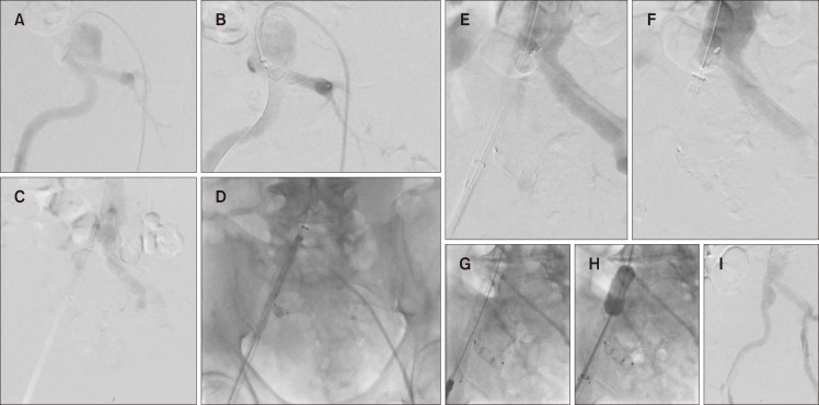
Fig. 3.
Intraoperative fluoroscopic image. (A-C) The introduction of a 28 cm long 18F sheath into the right common iliac artery. (D) The insertion of the 45 cm long 14F sheath using the peel-away showing both sheath tips matched at the same level. (E) The deployment of the first centimeter of the stent high into the aorta. (F) Advancing the 18F sheath to partially swallow back the deployed part of the endograft. (G-I) The completion of the deployment of the endograft demonstrating successful perfusion.

