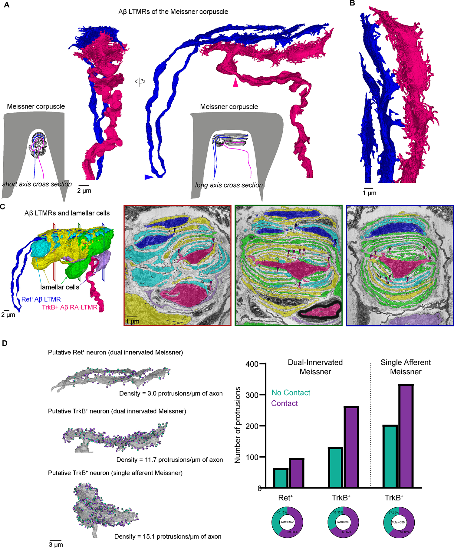
Figure 6. FIB-SEM reconstructions of two Meissner corpuscles reveal the density of axon protrusions to be a structural correlate of Aβ LTMR tactile sensitivity.

(A) 3D renderings of two Aβ LTMRs that innervate a Meissner corpuscle. Arrowheads indicate the termination of myelination.
(B) Higher magnification rendering of the terminal portion of the two Aβ LTMRs.
(C) Left, 3D renderings of the two Aβ LTMRs and four lamellar cells that form the Meissner corpuscle. Right, pseudo-colored images from the FIB-SEM volume at different depths across the corpuscle. Arrowheads show points where axon protrusions contact lamellar cells.
(D) Left, 3D renderings of the two axons from the dual-innervated corpuscle and of the axon from the single-innervated corpuscle. Teal dots indicate protrusions that terminate in the collagen (no contact). Purple dots indicate protrusions that contact lamellar cells. Right, quantification of protrusion terminals. See also Figure S6.
