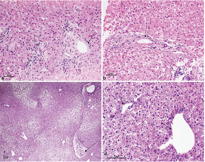
Supplementary Figure 2.

Obliterative portal venopathy. (a) Liver biopsy patient 4. Portal tract lacking a portal vein radicle (open arrow) next to a portal tract showing a portal vein radicle of reduced size (thin arrow). These two portal tracts are abnormally approximated to each other. (b) Liver biopsy patient 2. Portal tract showing a portal vein radicle of reduced size with a fibrous wall (arrow). (c) Liver biopsy patient 8. Thin fibrous septum connecting two portal tracts. The portal vein radicle shows a thick wall and a reduced size (arrow), while in other areas, the portal veins appear dilated. (d) Liver biopsy patient 8. Dilated portal veins herniating outside of the portal tracts in the periportal area indicating portal hypertension (arrows).
