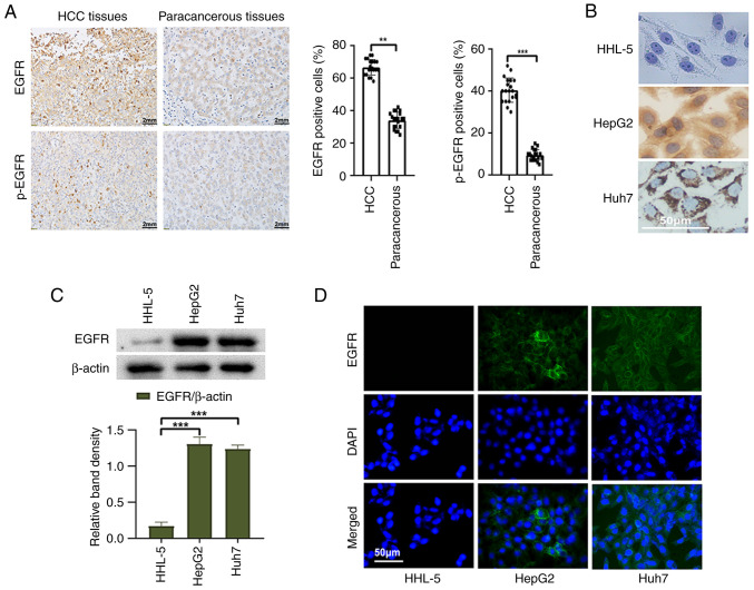
Figure 1.
Expression of EGFR in HCC tissues and liver cancer cells. (A) Representative images of immunohistochemical staining (magnification, ×400), and statistical analysis of the positive rates of EGFR and p-EGFR expression in HCC tissues and paracancerous tissues. (B) Immunocytochemistry staining, (C) western blotting and (D) indirect immunofluorescence was used to detect the expression of EGFR in normal liver cells and liver cancer cells. **P<0.01 and ***P<0.001. EGFR, epidermal growth factor receptor; HCC, hepatocellular carcinoma; p-, phosphorylated.

