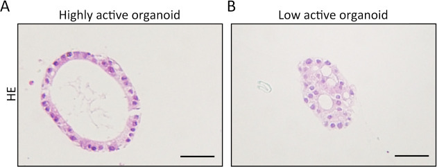
Figure 2.
Cellular vitality of organoids is evaluated by HE staining. (A) High-viability organoids from normal gastrointestinal epithelium exhibit a single-layer glandular structure with a polarity; (B) Cells of low-viability organoids are disordered with vacuoles appearance in the cytoplasm. The cellular polarity is lost. Scale bar 60 μm. HE, hematoxylin-eosin.

