Fig. 6.
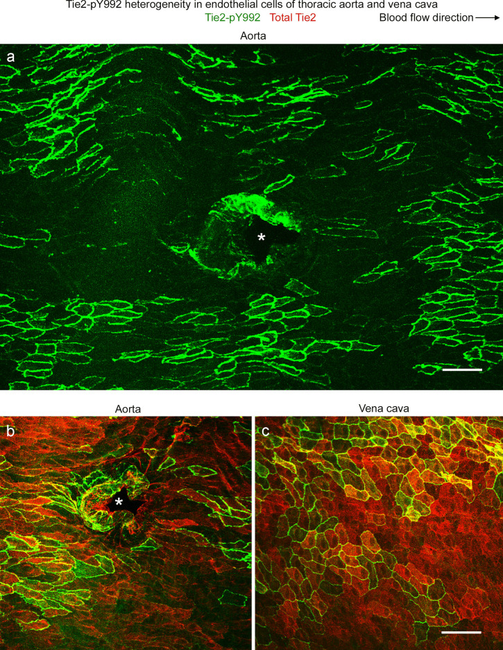
Tie2-pY992 heterogeneity in aorta and vena cava. a–c Confocal microscopic images showing the heterogeneous distribution of Tie2-pY922 (green) in endothelial cells of the thoracic aorta and vena cava of mice. The mosaic pattern of Tie2-pY992 consists of clusters of endothelial cells with strong staining surrounded by endothelial cells with little or no staining (a). Blood flow is left to right. b, c Broader distribution of overall Tie2 protein (red) than Tie2-pY992 (green) in the aorta (b) and vena cava (c). In both vessels, Tie2-pY992 staining is strongest at endothelial cell borders, whereas overall Tie2 is widespread. Asterisks mark intercostal artery ostia (a, b). Endothelial cells of the aorta are more elongated than those of the vena cava. Scale bars: 50 µm

