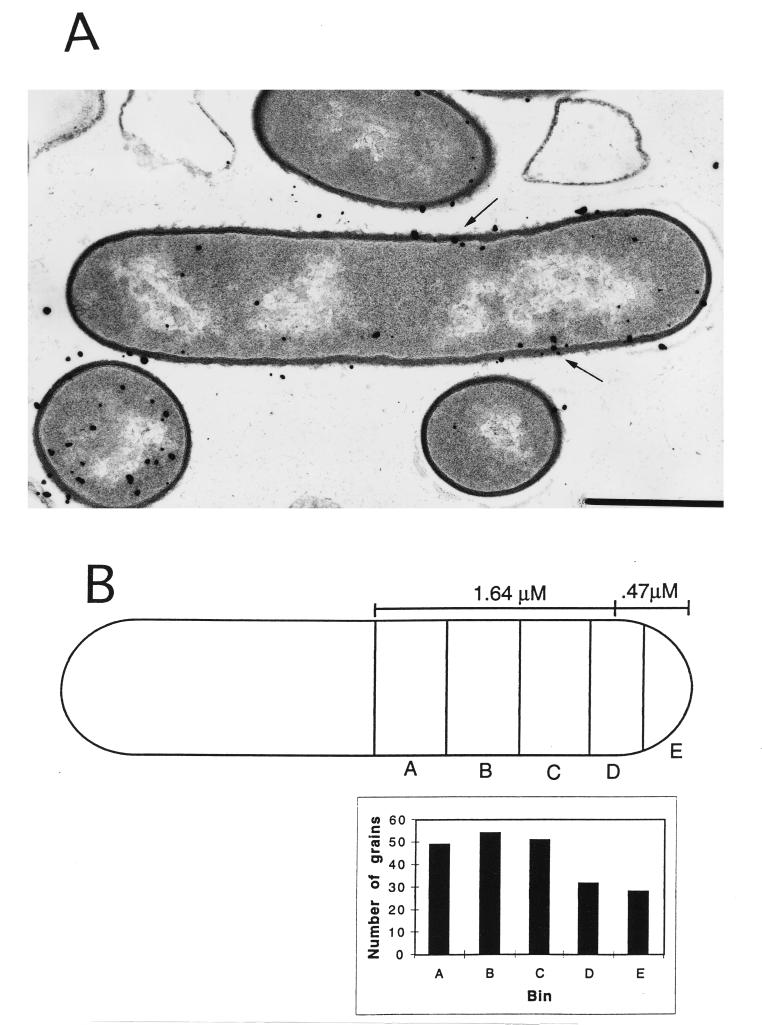
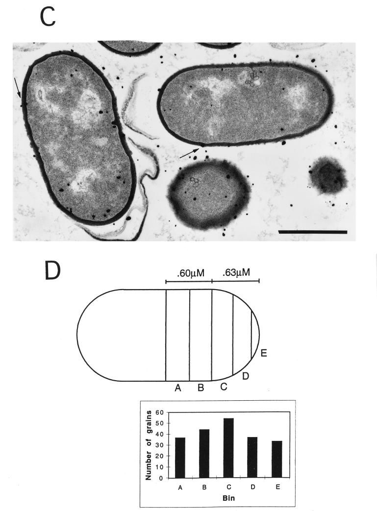
FIG. 3.
Electron microscopic autoradiography of wild-type and pbpA spores. The spores were heat shocked and inoculated into 2× YT medium with 4 mM l-alanine, as described in Materials and Methods. Sixty minutes after the initiation of spore germination, 500 μCi of [3H]N-acetyl-d-glucosamine was added and the culture was incubated for 2 min, followed by the addition of unlabeled N-acetyl-d-glucosamine to 1 mM. After 10 min of further incubation, the cells were harvested and processed for electron microscopic autoradiography, as described in Materials and Methods. Examples of wild-type (A) and pbpA (C) outgrowing spores are shown, with arrows indicating the labeled wall. The statistical analysis of silver grain distribution was done as described in Materials and Methods and is shown for the average wild-type (B) and pbpA (D) outgrowing spore after half of each cell was divided into 5 bins, with equal amounts of wall material in each bin. Bars, 1 μM.


