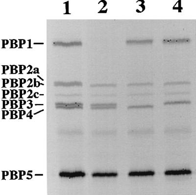
FIG. 4.

PBP profiles from membranes of various strains. Membranes were isolated from vegetative cells and labeled with FLU-C6-APA, as described in Materials and Methods. Approximately 10 μg of total membrane protein was run on sodium dodecyl sulfate–10% polyacrylamide gel electrophoresis for 4 h at 100 V, and labeled PBPs were visualized with a FluorimagerSI. The PBP pattern of wild-type cell membranes is like that previously found (lane 1) (18). Lane 1, PS832 (wild type); lane 2, PS2466 (pbpA ponA); lane 3, PS2467 (pbpA pbpC); lane 4, PS2468 (pbpA pbpD). The PBPs were designated as previously described (31).
