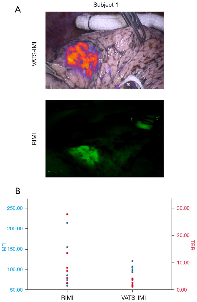
Figure 4.

RIMI compared to VIMI. (A) Intraoperative image of VIMI and RIMI in the same patient. (B) MFI of lesion and TBR measurements, stratified by imaging modality (RIMI vs. VIMI). Each blue dot on the scatter plot represents MFI (left y-axis) and red dot represents TBR (right y-axis) for an individual patient. There were no significant differences between groups. RIMI, robotic-assisted thoracic surgery intraoperative molecular imaging; VIMI, video-assisted thoracic surgery with intraoperative molecular imaging; MFI, mean fluorescence intensity; TBR, tumor-to-background ratio.
