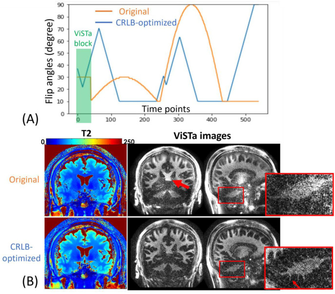
Figure 3.
(A) Original and CRLB-optimized flip angles (FA) of the ViSTa-MRF protocol. (B) T2 and ViSTa Comparisons between original ViSTa-MRF and CRLB-optimized ViSTa-MRF sequence. The zoom-in figures demonstrate that the CRLB-optimized ViSTa images exhibit higher SNR and better visualization of detailed structures in the cerebellum than the original ViSTa images.

