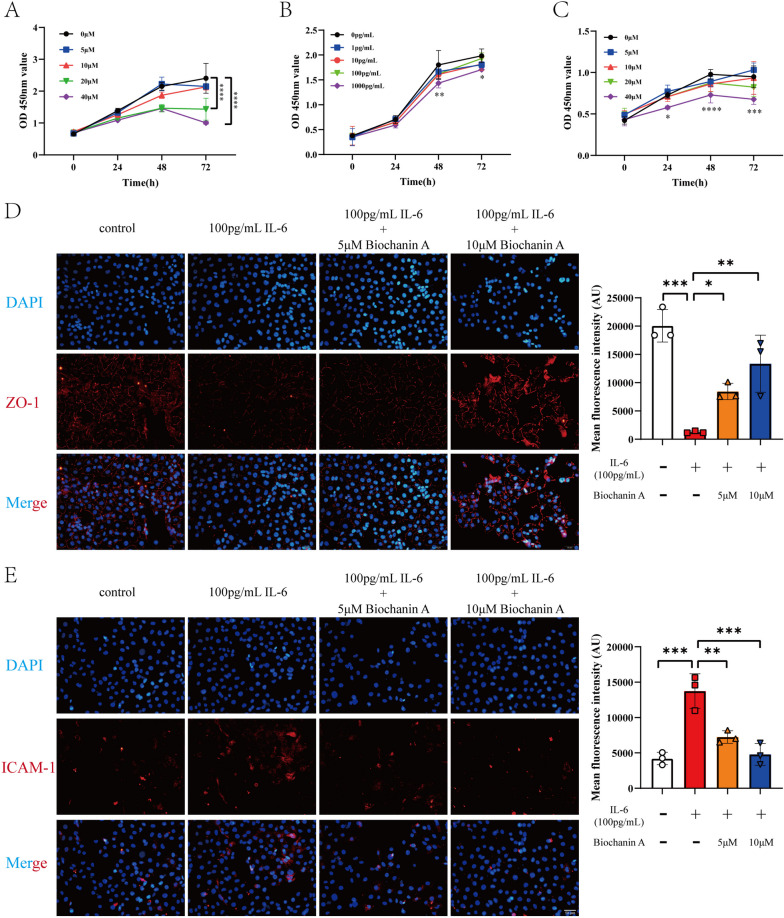
Fig. 2.
Biochanin A alleviates endothelial dysfunction induced by IL-6. A CCK8 result of different concentration of biochanin A showed that 0–10 μM biochanin A did not have toxic effect on HUVECs. (n = 3) B CCK8 result of different concentration of IL-6 showed that 0–100 pg/mL IL-6 did not have toxic effect on HUVECs. (n = 3) C CCK8 result of 100 pg/mL IL-6 combined with 0–10 μM biochanin A showed that the intervention did not have toxic effect on HUVECs. (n = 3) D The result of immunofluorescence showed that 100 pg/mL IL-6 caused the decreased expression of ZO-1 and biochanin A restored the expression of ZO-1. (n = 3) E The result of immunofluorescence showed that 100 pg/mL IL-6 caused the increased expression of ICAM-1 and biochanin A decreased the expression of ICAM-1. (n = 3) *p < 0.05, **p < 0.01, ***p < 0.001, ****p < 0.0001. #p < 0.05, 20 μM biochanin A compared with 0 μM biochanin A

