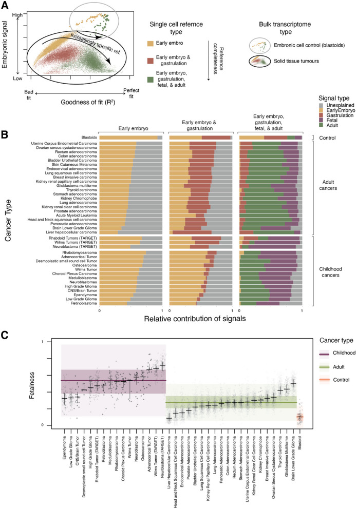
Fig. 1.
A Pan-cancer analysis of transformation state from pan-tissue single-cell reference atlases. Fit quality and embryonic signal of bulk transcriptomes with increasingly complete reference atlases: Fractional contribution of embryonic reference (y-axis, early embryo + gastrulation for full reference, early embryo otherwise) in explaining bulk transcriptomes (dots) as a function of goodness of fit (x-axis, pseudo R-squared) when fit using single cell reference consisting of cells from the early embryo (yellow), early embryo and gastrulation (dark red), or early embryo, gastrulation, foetal, and mature pan-tissue reference (green). Bulk cancer transcriptomes are circled in black and genuinely embryonal controls (blastoids) are circled in grey. B Relative contribution of references to explaining the bulk transcriptomes of a range of adult and childhood cancers: Average relative contribution of early embryo (yellow), gastrulation (dark red), foetal (purple), and adult (green) single cell reference populations in explaining bulk transcriptomes (y-axis) for different combinations of these references (x-axis, labels at top). Bulk transcriptomes are organised by source (labels on the right). C Childhood cancers have a stronger foetal contribution than adult cancers or control populations: Relative contribution of foetal reference (y-axis) in explaining bulk cancer transcriptomes (dots), when provided a complete set of the early embryo, gastrulation, foetal, and adult single-cell references. Bulk transcriptomes are split into childhood (purple), adult (green), and blastoid control (orange) and then by cancer type (x-axis). Distributions are summarised by median (horizontal lines), 1st and 3rd quartiles (horizontal lines for cancer types, shaded coloured areas for childhood/adult/control), and 1.5 times inter-quartile range (light-shaded areas)

