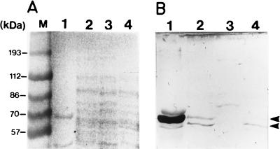
FIG. 5.
Western blot analysis. Cell extracts were subjected to polyacrylamide gel electrophoresis and transferred to nitrocellulose membranes as described in Materials and Methods. (A) Protein stained with Ponceau S; (B) Western blot carried out with rabbit polyclonal antibody raised against recombinant A. tumefaciens Pgm developed with peroxidase-conjugated antibody against rabbit immunoglobulin G (Dako). Lanes 1, commercial rabbit Pgm; lanes 2, A. tumefaciens wild-type extract; lanes 3, A. tumefaciens pgm mutant A5129 extract; lanes 4, A. tumefaciens glgB mutant A1120 extract. M, prestained molecular mass standards. Arrows on the right indicate the positions of Pgm proteins.

