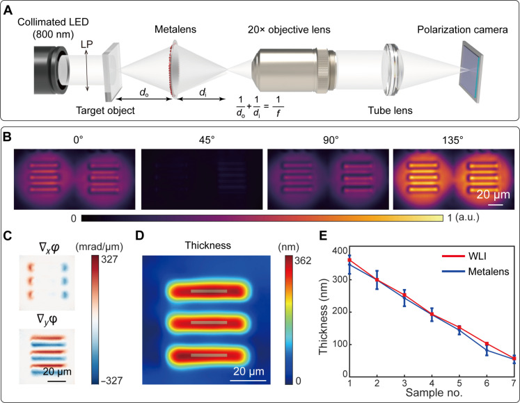
Fig. 4. Characterization of the metalens-assisted complex amplitude microscopy system.
(A) Schematic of the metalens-assisted complex amplitude microscopy system. (B) Captured shearing interference images with the polarization channel along 0°, 45°, 90°, and 135°, respectively. (C) Calculated phase gradients along the x and y direction, respectively. (D) Reconstructed thickness of the 361-nm-thick phase resolution target (group 6, element 1). (E) Comparison between the measured thickness of seven different phase targets by the proposed microscopy system and by a commercial WLI. Error bars represent SDs of the measured values.

