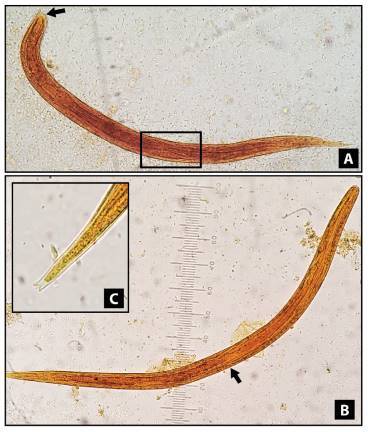
Figure 1. Strongyloides stercoralis larvae, stained with lugol solution, 400X. A) Rhabditform larvae showing a visible genital primordium (square) and short buccal canal (arrow). B) Filariform larvae displaying the union of the esophagus and the intestine in the middle of the larva (ratio 1:1). C) Filariform larvae with a detailed terminal part showing a notched tail.

