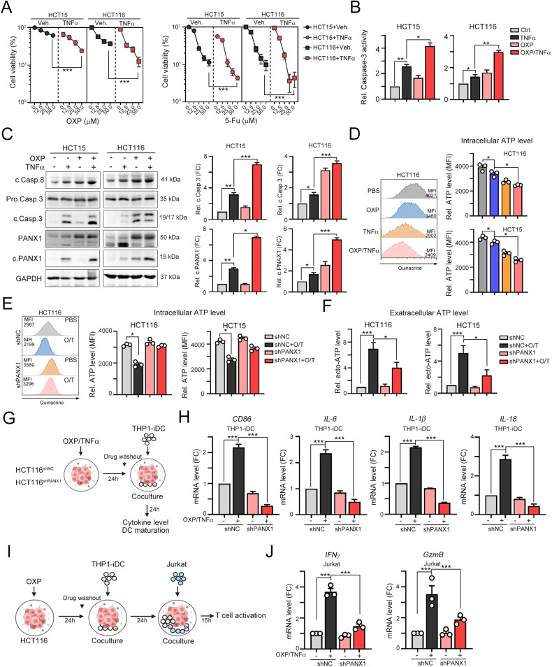
Fig. 3. TNFα enhances OXP-induced cell death and ATP release.
A HCT15 and HCT116 cells were treated with TNFα (50 ng/mL) and OXP or 5-Fu for 24 h. Cell viability was evaluated by CCK assay (n = 3). ***p < 0.001. Two-Way ANOVA test. B HCT15 and HCT116 cells were treated with TNFα (50 ng/mL) and OXP (25 μM) for 24 h. Caspase-3 activity was evaluated with a caspase-3 activity kit (n = 3). *p < 0.05 and **p < 0.01. One-Way ANOVA test. C HCT15 and HCT116 cells were treated with TNFα (50 ng/mL) and OXP (25 μM) for 24 h. The cleavage of caspase 8, caspase-3 and PANX1 was evaluated by western blotting (n = 3). *p < 0.05, **p < 0.01 and ***p < 0.001. One-Way ANOVA test. D After treatment with TNFα and OXP for 6 h, the intracellular ATP content was examined by flow cytometry (n = 3). *p < 0.05. One-Way ANOVA test. E HCT15shNC and HCT15shPANX1 cells were treated with TNFα (50 ng/mL) and OXP (25 μM) for 6 h. The intracellular ATP content was examined by flow cytometry (n = 3). *p < 0.05. One-Way ANOVA test. Similar experiments were carried out in HCT116 cells. F HCT15shNC and HCT15shPANX1 cells were treated with TNFα (50 ng/mL) and OXP (25 μM) for 6 h. The extracellular ATP content was examined with a luminescent ATP detection kit (n = 3). *p < 0.05 and ***p < 0.001. One-Way ANOVA test. Similar experiments were carried out in HCT116 cells. G Schematic diagram of the DC maturation analysis. HCT116shNC and HCT116shPANX1 cells were treated with TNFα (50 ng/mL) and OXP (25 μM) for 24 h. Then, these cells were cocultured with THP1- iDCs for 24 h. H The mRNA levels of the DC maturation marker CD86 and the proinflammatory cytokines IL-6, IL-1β and IL-18 in THP-iDCs that were cocultured with vehicle- or OXP/TNFα-treated HCT116shNC and HCT116shPANX1 cells were evaluated by qRT‒PCR (n = 3). ***p < 0.001. One-Way ANOVA test. I HCT116shNC and HCT116shPANX1 cells were treated with TNFα (50 ng/mL) and OXP (25 μM) for 24 h. Then, these cells were cocultured with THP1-iDCs for 24 h and Jurkat T cells for 15 h. J The mRNA levels of the cytotoxic cytokines IFNγ and GzmB in Jurkat T cells that were cocultured with THP-iDCs and vehicle- or OXP/TNFα-treated HCT116shNC/HCT116shPANX1 cells was evaluated by qRT‒PCR (n = 3). ***p < 0.001. One-Way ANOVA test.

