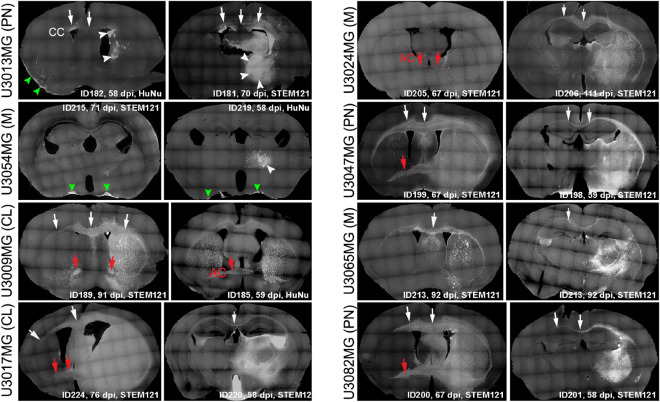
Figure 1.
In vivo growth patterns of tumors derived from eight GSC lines. Whole coronal sections stained with STEM121 or HuNu antibodies to localize human glioblastoma cells, followed by imaging using the Leica SP8 microscope tile scan function. Animal ID and tumor age in days post injection (dpi) are indicated. CC corpus callosum (white arrows), AC anterior commissure (red arrows); Subarachnoid space (green arrowheads); Solid tumor mass (white arrowheads). GBM molecular subtypes of GSC lines are indicated: CL classical, M mesenchymal, PN proneural.

