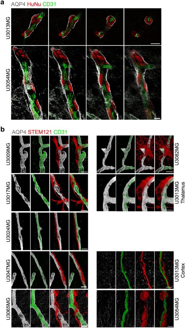
Figure 4.

Perivascular invasion and displacement of astrocyte end-feet by glioma cells. (a) U3013MG and U3054MG glioma cells in perivascular space (between blood vessel and astrocyte end-feet). Coronal sections were stained with HuNu (red, human glioma cells), AQP4 (white, astrocyte end-feet) and CD31 (green, blood vessels) antibodies and z-stacks of selected regions were taken by Leica confocal microscope using 63 × objective. Scale bar, 20 µm. (b) Interaction of human glioma cells (STEM121, red) with astrocyte end-feet and blood vessels. Each image represents a snapshot of a 3D image. Scale bar, 10 µm.
